. 2014 Aug;88(16):8813-25.
doi: 10.1128/JVI.00992-14. Epub 2014 May 28.

Virus particle release from glycosphingolipid-enriched microdomains is essential for dendritic cell-mediated capture and transfer of HIV-1 and henipavirus

Affiliations

Virus particle release from glycosphingolipid-enriched microdomains is essential for dendritic cell-mediated capture and transfer of HIV-1 and henipavirus

Hisashi Akiyama et al. J Virol. 2014 Aug.

Abstract

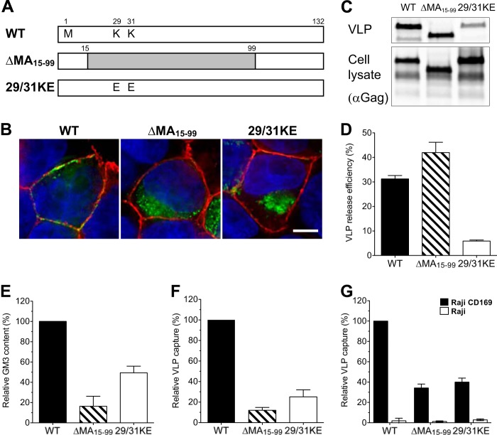
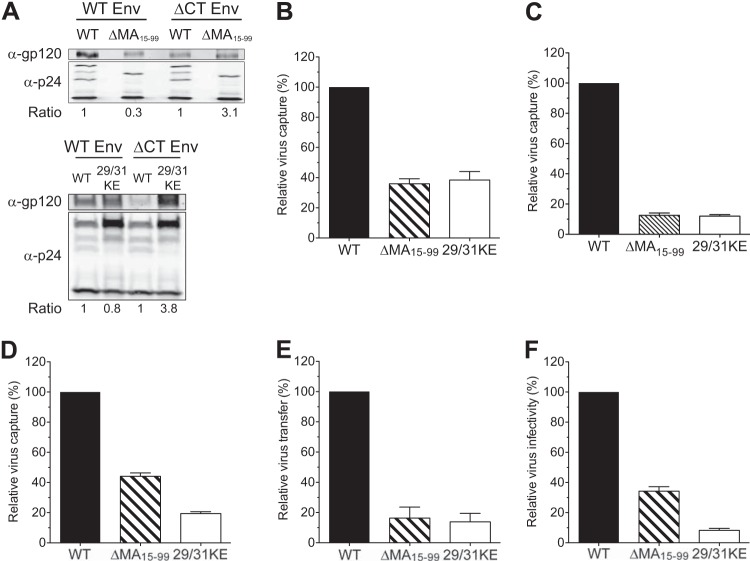
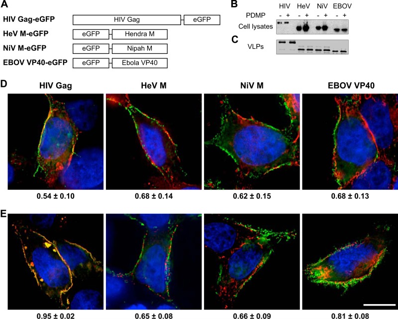
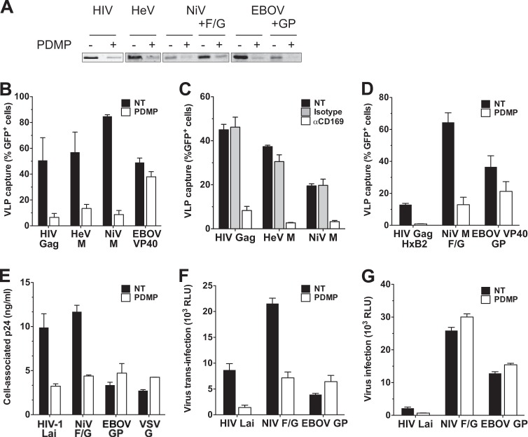
Human immunodeficiency virus type 1 (HIV-1) exploits dendritic cells (DCs) to promote its transmission to T cells. We recently reported that the capture of HIV-1 by mature dendritic cells (MDCs) is mediated by an interaction between the glycosphingolipid (GSL) GM3 on virus particles and CD169/Siglec-1 on MDCs. Since HIV-1 preferentially buds from GSL-enriched lipid microdomains on the plasma membrane, we hypothesized that the virus assembly and budding site determines the ability of HIV-1 to interact with MDCs. In support of this hypothesis, mutations in the N-terminal basic domain (29/31KE) or deletion of the membrane-targeting domain of the HIV-1 matrix (MA) protein that altered the virus assembly and budding site to CD63(+)/Lamp-1-positive intracellular compartments resulted in lower levels of virion incorporation of GM3 and attenuation of virus capture by MDCs. Furthermore, MDC-mediated capture and transmission of MA mutant viruses to T cells were decreased, suggesting that HIV-1 acquires GSLs via budding from the plasma membrane to access the MDC-dependent trans infection pathway. Interestingly, MDC-mediated capture of Nipah and Hendra virus (recently emerged zoonotic paramyxoviruses) M (matrix) protein-derived virus-like particles that bud from GSL-enriched plasma membrane microdomains was also dependent on interactions between virion-incorporated GSLs and CD169. Moreover, capture and transfer of Nipah virus envelope glycoprotein-pseudotyped lentivirus particles by MDCs were severely attenuated upon depletion of GSLs from virus particles. These results suggest that GSL incorporation into virions is critical for the interaction of diverse enveloped RNA viruses with DCs and that the GSL-CD169 recognition nexus might be a conserved viral mechanism of parasitization of DC functions for systemic virus dissemination.

Importance: Dendritic cells (DCs) can capture HIV-1 particles and transfer captured virus particles to T cells without establishing productive infection in DCs, a mechanism of HIV-1 trans infection. We have recently identified CD169-mediated recognition of GM3, a host-derived glycosphingolipid (GSL) incorporated into the virus particle membrane, as the receptor and ligand for the DC-HIV trans infection pathway. In this study, we have identified the matrix (MA) domain of Gag to be the viral determinant that governs incorporation of GM3 into HIV-1 particles, a previously unappreciated function of the HIV-1 MA. In addition, we demonstrate that the GSL-CD169-dependent trans infection pathway is also utilized as a dissemination mechanism by henipaviruses. GSL incorporation in henipaviruses was also dependent on the viral capsid (M) protein-directed assembly and budding from GSL-enriched lipid microdomains. These findings provide evidence of a conserved mechanism of retrovirus and henipavirus parasitization of cell-to-cell recognition pathways for systemic virus dissemination.


Figures

FIG 1
FIG 1
MA mutant Gag-eGFP VLPs bud from intracellular membranes, and capture of HIV-1 Gag-eGFP VLPs by mature DCs is MA dependent. (A) Construction of HIV-1 matrix mutants. (B) Localization of eGFP fusion proteins in transfected HEK293T cells stained with CTxB-Alexa 594 for GM1 (red) and with DAPI for the nucleus (blue). Bar, 5 μm. (C) Western blot analysis of VLPs produced from transfected HEK293T cells was performed by probing with an anti-p24Gag monoclonal antibody. (D) Efficiency of VLP release from HEK293T cells. The amount of p24Gag in transfected HEK293T cell lysates and supernatants was quantified, and VLP release efficiencies were calculated [release efficiency = amount of p24Gag in the supernatant/(amount of p24Gag in the cell lysates + amount of p24Gag in the supernatant)]. The data shown are the means of triplicate experiments ± SDs. WT, wild-type Gag-eGFP; Δ15-99, ΔMA15-99-eGFP; 29/31KE, 29/31KE-eGFP. (E) The amount of VLP-associated GM3 was quantified by immunofluorescence staining. The value was normalized to that of wild-type Gag-eGFP. The experiment was performed on at least two independent VLP preparations, and the data are reported as the means ± SEMs of a representative experiment. (F) Mature DCs were challenged with VLPs and analyzed for GFP-positive cells by FACS. The percentage of GFP-positive DCs was normalized to that of wild-type Gag-eGFP VLPs. The results represent the means ± SEMs from four independent experiments performed on DCs from four different donors. (G) Raji-CD169 cells or Raji cells were challenged with VLPs, washed, and analyzed for GFP-positive cells. The value was normalized to that of wild-type Gag-eGFP capture by Raji-CD169 cells. The experiment was performed in triplicate at least two independent times, and the data from a representative experiment are shown as the means ± SDs.
FIG 2
FIG 2
HIV-1 MA mutants are severely attenuated in DC-mediated capture and trans infection of T cells. (A) Quantitative Western blotting of incorporation of wild-type gp120 or gp120 with a cytoplasmic tail deletion in virus particles with wild-type (WT), ΔMA15-99, or 29/31KE matrix proteins. The band intensity was quantified, and the ratio of the amount of gp120 to the total amount of Gag (p24Gag + Pr55Gag) in ΔMA15-99 or 29/31KE virions was normalized to that observed with wild-type Gag containing wild-type Env or ΔCT Env. (B to D) Mature DCs were challenged with NL4-3 viruses containing intact Env (B), NL4-3 viruses containing ΔCT Env (C), or NL4-3 viruses without Env (ΔEnv) (D) and encoding the wild-type, ΔMA15-99, or 29/31KE matrix protein. Cells were washed and lysed for cell-associated p24Gag measurement, and the results were normalized to those observed with viruses encoding wild-type Gag. The data shown are the means ± SEMs of three (B and D) or nine (C) independent experiments with DCs from different donors. (E) Mature DCs were challenged with NL4-3ΔCT viruses encoding the wild-type, ΔMA15-99, or 29/31KE matrix proteins and washed, and DC-to-T cell (MT4) trans infection was quantified. The values were normalized to those for NL4-3ΔCT (wild type). (F) In parallel, cell-free infection of MT4 cells was examined. The data shown are the means ± SEMs of three independent experiments with DCs from three different donors (E) or of four independent experiments performed in triplicate (F).
FIG 3
FIG 3
Paramyxovirus and filovirus VLPs bud from GSL-enriched lipid microdomains at the plasma membrane. (A) Constructions of HeV M, NiV M, and EBOV VP40 fusion proteins. (B and C) Western blot analysis of eGFP fusion proteins in transfected HEK293T cell lysates (B) and VLPs in the supernatants (C) in the presence or absence of PDMP. (D) Localization of eGFP fusion proteins in transfected HEK293T cells stained with CTxB for GM1 (red) and with DAPI for the nucleus (blue). The values at the bottom are Pearson's colocalization coefficients of the means ± SDs. (E) Localization of Gag-mCherry and the indicated eGFP fusion proteins in transfected HEK293T cells stained with DAPI (blue). The values at the bottom are Pearson's colocalization coefficients of the means ± SDs. Bar, 10 μm.
FIG 4
FIG 4
Capture of Hendra and Nipah virus VLPs by DCs is dependent on GSL-CD169 interaction. (A) VLPs derived from transfected HEK293T cells in the presence or absence of PDMP were immunoprecipitated with CTxB. Immunoprecipitated VLPs were probed for GFP expression via quantitative Western blot analysis. (B) Mature DCs were incubated with VLPs containing equal amounts of GFP produced in the presence or absence of PDMP, washed, and analyzed for GFP-positive cells by flow cytometry. (C) Mature DCs were untreated or incubated with isotype control antibody or anti-CD169 blocking antibody, challenged with VLPs, washed, and analyzed for GFP-positive cells by flow cytometry. (D) Mature DCs were incubated with VLPs pseudotyped with the indicated envelope glycoproteins produced in the presence or absence of PDMP and analyzed for GFP-positive cells by flow cytometry. (E) Mature DCs were challenged with pseudotyped HIV-1 vectors produced in the presence or absence of PDMP and washed, and the amount of cell-associated p24Gag was measured. (F) Mature DCs were incubated with pseudotyped HIV-1 vectors produced in the presence or absence of PDMP, washed, and cocultured with U373/CD4/CXCR4 cells, and the cells were lysed at 2 days postinfection to measure the luciferase activity in lysates. (G) U373/CD4/CXCR4 cells were infected with cell-free pseudotyped HIV-1 vectors produced in the presence or absence of PDMP, and cells were lysed at 2 days postinfection to measure the luciferase activity in cell lysates as a measure of virus infection. The data shown are the means ± SEMs of three independent experiments with DCs from three different donors. NT, not treated; RLU, relative light units.

References

    1. Hladik F, McElrath MJ. 2008. Setting the stage: host invasion by HIV. Nat. Rev. Immunol. 8:447–457. 10.1038/nri2302 - DOI - PMC - PubMed
    1. Li Q, Estes JD, Schlievert PM, Duan L, Brosnahan AJ, Southern PJ, Reilly CS, Peterson ML, Schultz-Darken N, Brunner KG, Nephew KR, Pambuccian S, Lifson JD, Carlis JV, Haase AT. 2009. Glycerol monolaurate prevents mucosal SIV transmission. Nature 458:1034–1038. 10.1038/nature07831 - DOI - PMC - PubMed
    1. Hladik F, Sakchalathorn P, Ballweber L, Lentz G, Fialkow M, Eschenbach D, McElrath MJ. 2007. Initial events in establishing vaginal entry and infection by human immunodeficiency virus type-1. Immunity 26:257–270. 10.1016/j.immuni.2007.01.007 - DOI - PMC - PubMed
    1. Hu J, Gardner MB, Miller CJ. 2000. Simian immunodeficiency virus rapidly penetrates the cervicovaginal mucosa after intravaginal inoculation and infects intraepithelial dendritic cells. J. Virol. 74:6087–6095. 10.1128/JVI.74.13.6087-6095.2000 - DOI - PMC - PubMed
    1. Spira AI, Marx PA, Patterson BK, Mahoney J, Koup RA, Wolinsky SM, Ho DD. 1996. Cellular targets of infection and route of viral dissemination after an intravaginal inoculation of simian immunodeficiency virus into rhesus macaques. J. Exp. Med. 183:215–225. 10.1084/jem.183.1.215 - DOI - PMC - PubMed

Publication types

MeSH terms

Substances

LinkOut - more resources