. 2014 Aug;88(16):8843-52.
doi: 10.1128/JVI.00424-14. Epub 2014 May 28.

Activation of c-jun N-terminal kinase upon influenza A virus (IAV) infection is independent of pathogen-related receptors but dependent on amino acid sequence variations of IAV NS1

Affiliations

Activation of c-jun N-terminal kinase upon influenza A virus (IAV) infection is independent of pathogen-related receptors but dependent on amino acid sequence variations of IAV NS1

Wolfgang Nacken et al. J Virol. 2014 Aug.

Abstract

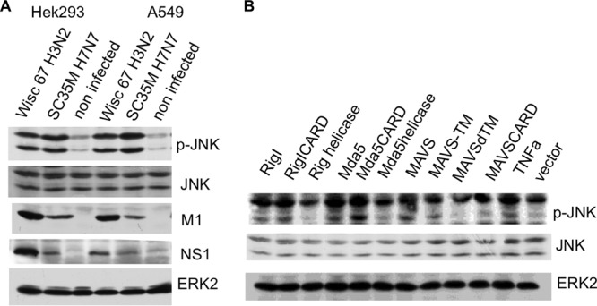
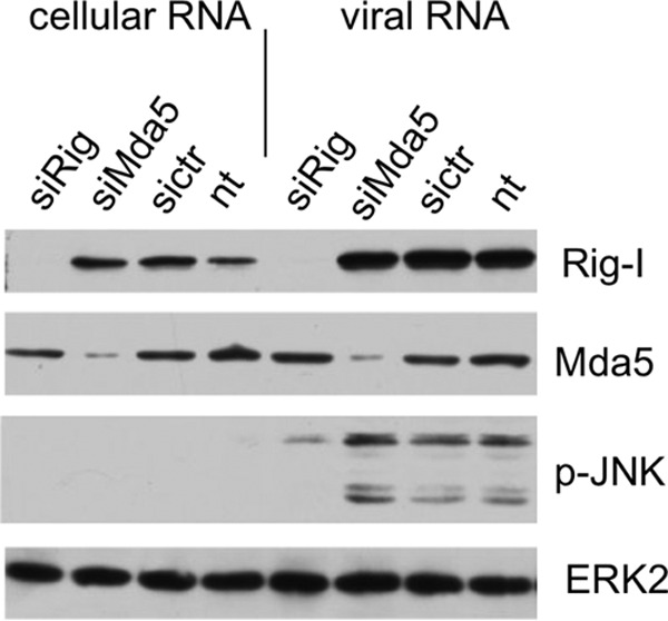
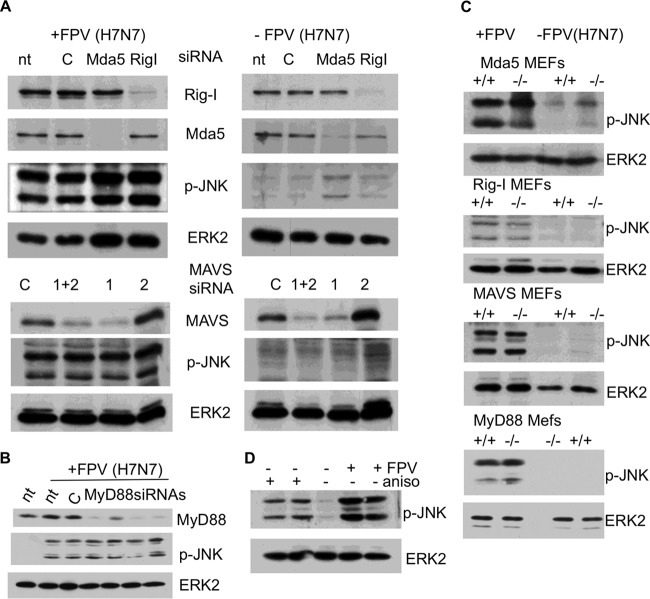
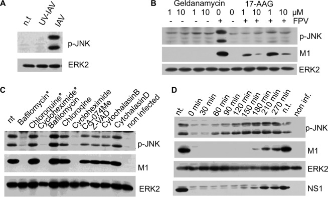
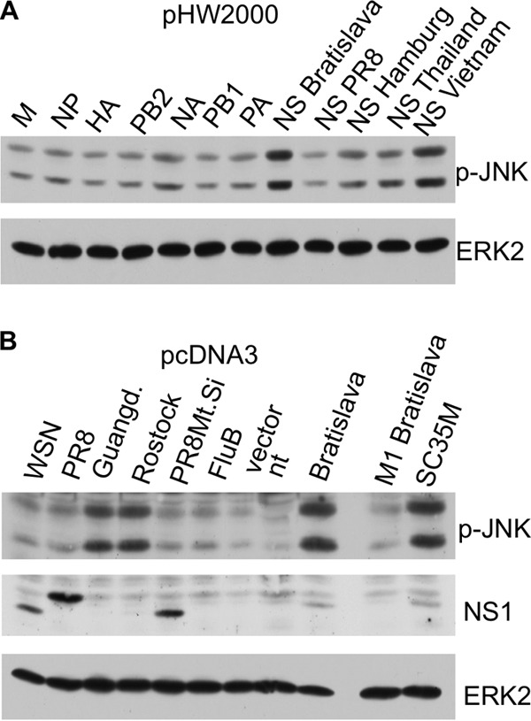
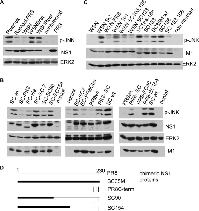
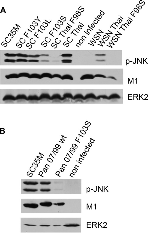
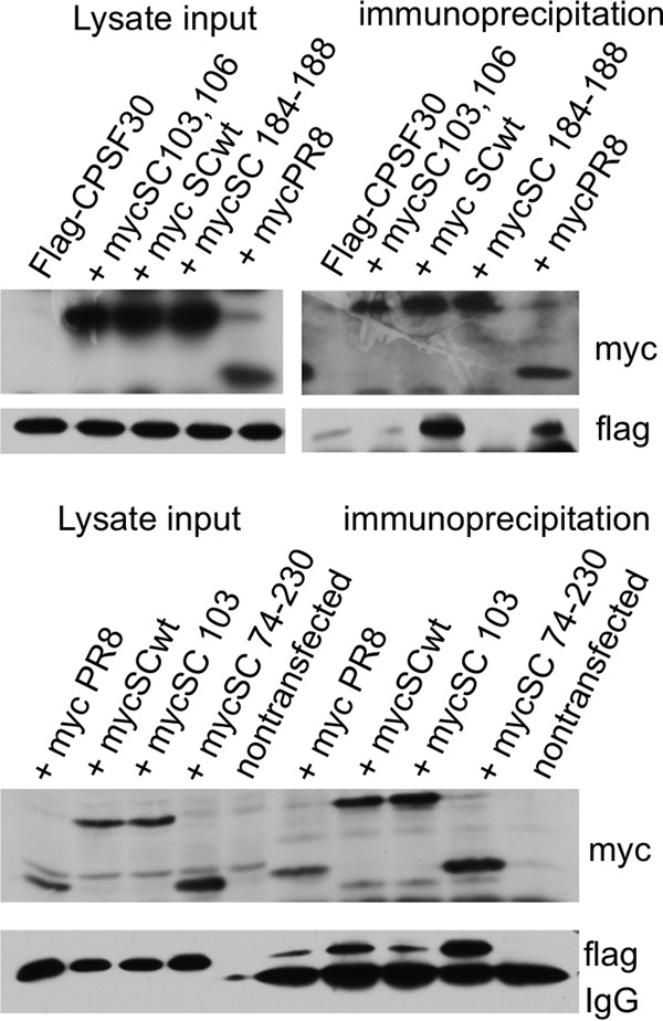
A hallmark cell response to influenza A virus (IAV) infections is the phosphorylation and activation of c-jun N-terminal kinase (JNK). However, so far it is not fully clear which molecules are involved in the activation of JNK upon IAV infection. Here, we report that the transfection of influenza viral-RNA induces JNK in a retinoic acid-inducible gene I (RIG-I)-dependent manner. However, neither RIG-I-like receptors nor MyD88-dependent Toll-like receptors were found to be involved in the activation of JNK upon IAV infection. Viral JNK activation may be blocked by addition of cycloheximide and heat shock protein inhibitors during infection, suggesting that the expression of an IAV-encoded protein is responsible for JNK activation. Indeed, the overexpression of nonstructural protein 1 (NS1) of certain IAV subtypes activated JNK, whereas those of some other subtypes failed to activate JNK. Site-directed mutagenesis experiments using NS1 of the IAV H7N7, H5N1, and H3N2 subtypes identified the amino acid residue phenylalanine (F) at position 103 to be decisive for JNK activation. Cleavage- and polyadenylation-specific factor 30 (CPSF30), whose binding to NS1 is stabilized by the amino acids F103 and M106, is not involved in JNK activation. Conclusively, subtype-specific sequence variations in the IAV NS1 protein result in subtype-specific differences in JNK signaling upon IAV infection.

Importance: Influenza A virus (IAV) infection leads to the activation or modulation of multiple signaling pathways. Here, we demonstrate for the first time that the c-jun N-terminal kinase (JNK), a long-known stress-activated mitogen-activated protein (MAP) kinase, is activated by RIG-I when cells are treated with IAV RNA. However, at the same time, nonstructural protein 1 (NS1) of IAV has an intrinsic JNK-activating property that is dependent on IAV subtype-specific amino acid variations around position 103. Our findings identify two different and independent pathways that result in the activation of JNK in the course of an IAV infection.


Figures

FIG 1
FIG 1
JNK is activated upon IAV infection and by overexpression of RIG-I-like receptor. (A) A549 and HEK293 cells were infected with the human IAV strain A/Wisconsin (Wisc)/67/2005 (H3N2) and avian IAV SC35M (H7N7) at an MOI of 5. After 4 h, cells were harvested and lysates were subjected to an SDS-PAGE and subsequently analyzed by Western blotting with antibodies against the phosphorylated active form of JNK1 and JNK2 (p-JNK), JNK, NS1, and M1. ERK2 served as a loading control. (B) Cells were transfected with expression plasmids as indicated. Cell lysates were harvested 24 h after transfection and analyzed by Western blotting with an anti-phospho-JNK antibody (p-JNK); ERK2 blots served as a loading control. RIG-I, Mda5, and MAVS, plasmids expressing wild-type proteins; RIG-I CARD, RIG-I CARD domain; RIG-I helicase, RIG-I helicase domain; MAVS-TM, MAVS aa 1 to 510 with the transmembrane domain of bcl-xl added; MAVSdTM, MAVS aa 1 to 510 with the transmembrane domain deleted; MAVS CARD, MAVS aa 1 to 98; TNFa, tumor necrosis factor alpha-treated cells.
FIG 2
FIG 2
The activation of JNK upon viral-RNA transfection is RIG-I dependent. A549 cells were transfected with siRNA targeting RIG-I (siRig) or Mda5 (siMda5). Subsequently, the cells were transfected with viral RNA or cellular RNA, harvested after 4 h, and analyzed by Western blotting. sictr, control siRNA; nt, not transfected with siRNA.
FIG 3
FIG 3
IAV infection-mediated JNK activation is independent of RIG-I like receptors and of MyD88-dependent TLRs. (A) A549 cells were transfected with siRNA targeting RIG-I, Mda5, and MAVS, as indicated. Cells were infected at an MOI of 5 with A/FPV/Bratislava/79 (FPV [H7N7]) 48 h after siRNA transfection. Subsequently, cell lysates were harvested 4 h postinfection and analyzed by Western blotting. p-JNK, Western blot with an anti phospho-JNK antibody; ERK2, control blot using anti-ERK2 antibody; Mda5, RIG-I, and MAVS, Western blots using antibodies specific to Mda5, RIG-I, and MAVS, respectively; nt, nontreated; C, control siRNA; 1 and 2, two different siRNAs targeting MAVS (MAVS siRNA 2 was not effective in suppressing MAVS expression); 1+2, MAVS siRNA 1 and 2 mixed. (B) Identical experiments were conducted using four different MyD88 siRNAs. nt, nontreated; C, control siRNA. (C) To further exclude any role of these genes in IAV-mediated JNK activation, MEFs deficient (−/−) for RIG-I, Mda5, MAVS, or MyD88 and the corresponding wild-type MEFs (+/+) were either infected with FPV/Bratislava/79 (FPV) (MOI of 5) or left uninfected. Cell lysates were harvested 4 h postinfection and analyzed by Western blotting using an anti-phospho-JNK antibody; ERK2 blots served as a loading control (ERK2). (D) Chicken DF-1 cells are reported to be devoid of a functional RIG-I (27). (A) DF-1 cells were infected with A/FPV/Bratislava/79 (FPV [H7N7]) and analyzed by Western blotting using anti-phospho-JNK antibodies. As a positive control, DF-1 cells were incubated with anisomycin (aniso), which is a potent JNK activator. As indicated, the anisomycin-positive control experiment and the infection experiment were performed in duplicate.
FIG 4
FIG 4
The expression of functional viral proteins is needed for IAV-induced JNK activation. (A) A/FPV/Bratislava/79 (H7N7) (IAV) was inactivated by UV light. Cells were infected with native and UV-treated IAV and analyzed by Western blotting. (B) Cells were infected with A/FPV/Bratislava/79 (FPV [H7N7]), subsequently incubated with inhibitors of the heat shock protein hsp90 (geldanamycin, 17-AAG), and analyzed via Western blotting. (C) Cells were incubated with FPV/Bratislava/79 (H7N7) for 30 min, and subsequently inhibitors were added as indicated. After 4 h, cells were harvested and analyzed via Western blotting. nt, nontreated. * indicates samples that were additionally treated with the indicated inhibitor 20 min before infection. (D) Cells were infected with FPV/Bratislava/79 (H7N7) (MOI of 5) by incubating them for 30 min with the virus-containing supernatant. After removal of the supernatant, cycloheximide was added at the indicated time points postinfection and harvested 270 min postinfection. nt, nontreated; non inf., noninfected.
FIG 5
FIG 5
IAV NS1 expression induces JNK activation. (A) pHW2000- and pMPccdB-based plasmids encoding the eight genomic segments of A/FPV/Bratislava/79 were transfected into A549 cells. Additionally, the pHW2000 plasmids containing NS segments of A/Puerto Rico/8/34 (H1N1) (PR8), A/Hamburg/4/09 (H1N1) (Hamburg), A/Thailand/1(Kan-1)/04 (H5N1) (Thailand), and A/Vietnam/1203/04 (H5N1) (Vietnam) were transfected into cells. After 24 h, cells were harvested and analyzed by Western blotting. M, matrix protein; NP, nuclear protein; HA, hemagglutinin; PB2, polymerase B2; NA, neuraminidase; PB1, polymerase B1; PA, polymerase A. (B) pcDNA3 plasmids expressing NS1s from IAVs of various subtypes were transfected into cells, and phosphorylation of JNK was analyzed by Western blotting. In the cases of the Guangdong and Rostock viruses, either the monoclonal NS1 antibody could not detect the subtype-specific NS1 protein or expression was too low to be detected. Guangd., NS1 from A/Goose/Guangdong/3/97 (H5N1); Rostock, A/FPV/Rostock/34 (H7N1); WSN, A/WSN/1933 (H1N1); PR8, A/Puerto Rico/8/34 (H1N1); FluB, influenza B virus; PR8Mt.Si, A/Puerto Rico/8/34/Mount Sinai (H1N1); Bratislava, NS1 from FPV/Bratislava/79 (H7N7); M1 Bratislava, M1 from FPV/Bratislava/79 (H7N7); SC35M, SC35M (H7N7).
FIG 6
FIG 6
JNK activation depends on amino acid position 103 of the SCM35M NS1 protein. (A) Cells were infected with IAV A/FPV/Rostock/34 (Rostock), A/WSN/1933 (WSN), and A/Puerto Rico/8/34 (PR8) and with reassortant viruses, such as A/FPV/Rostock/34 containing an NS segment from A/Puerto Rico/8 (RostockPR8) and IAV WSN/1933 containing an NS segment from A/FPV/Bratislava/79 (WSNBrat) or from A/FPV/Rostock/34 (WSNRost). Subsequently, cell lysates were analyzed by Western blotting as described in the text. (B) Cells were infected with recombinant IAVs for 4 h and analyzed by Western blotting. SC wt, wild-type SC35M; SC-PR8, SC35M containing the NS segment from A/Puerto Rico/8; SC-Ha, SC35M containing the NS segment from A/Hamburg/4/09; SC-SC 7, SC35M with an SC35M NS segment mutated at seven amino acid positions into the corresponding amino acids of PR8, namely, S3P, F22V, I81M, S114P, P215T, R225G, and E228R; SC-SC90, SC35M containing a chimeric NS segment including amino acids 1 to 90 of SC35M, followed by part of A/Puerto Rico/8, and with the C-terminal amino acid positions 215, 224, and 227 mutated into the corresponding amino acids of SC35M; SC-SC154, SC35M containing a chimeric NS segment that is the same as SC90 except that amino acids 1 to 154 are those encoded by SC35M; PR8wt, IAV Puerto Rico/8/34 containing the NS segment of SC35M; SC-PR8Cter, SC35M IAV containing the NS segment of PR8 mutated at amino acid positions 215, 224, and 227 into the corresponding amino acids of SC35M; PR8-SC, PR8 containing the NS segment of SC35M; PR8-SC90, PR8 with the NS segment of SC90; PR8-SC154, PR8 with the NS segment of SC154. (Right) Schematic representation of chimeric NS SC90 and SC154 segments (see panel D). (C) Cells were infected with recombinant IAVs and analyzed as described above. WSN, A/WSN/1933; WSN SC, WSN with the NS segment of SC35M; WSN PR8, WSN with the NS segment of PR8; WSN 101, WSN with an NS segment containing H101D; WSN SC103, 106, WSN with SC35M NS F103S M106I; WSN SC103, WSN with SC35M NS1 F103S; SC184-188, SC35M with NS with a mutation of GLEWN at positions 184 to 188 of RFKRY; SC103, SC35M with NS F103S; SC106, SC35M with NS M106I; SC103, 106, SC35M with NS F103S M106I. (D) Schematic representation of the chimeric NS1 proteins described for panel B.
FIG 7
FIG 7
Serine at position 103 of NS1 is not compatible with JNK activation. Cells were infected with recombinant IAVs for 4 h and analyzed by Western blotting. (A) SC35M, wild-type SC35M; SC F103Y, SC35M encoding NS1 F103Y; SCF103L, SC35M encoding NS1 F103L; SC F103S, SC35M encoding NS1 F103S; SC35 Thai, SC35M encoding the NS segment of A/Thailand/1 (KAN-1); SC Thai F98S, SC35M encoding the NS segment of A/Thailand/1 (Kan-1) with F98S; WSN, A/WSN/1933; WSN Thai, A/WSN/1933 encoding the NS segment of Thailand/1 (KAN-1); WSN Thai F98S, A/WSN/1933 encoding the NS segment of Thailand/1 (KAN-1) with F98S. (B) Pan 07/99 wt, wild-type A/Panama 2007/99; Pan 07/99 F103S, A/Panama 2007/99 encoding the NS1 F103S mutant.
FIG 8
FIG 8
Cells were transfected with Flag-tagged CPSF30 and various myc-tagged NS1 expression constructs. Lysates were immunoprecipitated using a monoclonal anti-myc antibody. Flag-CPSF30, Flag-tagged CPSF30; mycSC103, 106, 6×-myc-tagged SC35M NS1 F103S M106I; myc SCwt, 6×-myc-tagged SC35M NS1; mycSC 184-188, 6×-myc-tagged SC35M NS1 with a mutation of GLEWN at positions 184 to 188 to RFKRY; mycPR8, 1×-myc-tagged PR8 NS1; mycSC103, 6×-myc-tagged SC35M NS1 F103S; mycSC 74-230, 6×-myc-tagged NS1 with amino acid positions 74 to 230 (effector domain).

Similar articles

Cited by

References

    1. Eliopoulos AG, Blake SM, Floettmann JE, Rowe M, Young LS. 1999. Epstein-Barr virus-encoded latent membrane protein 1 activates the JNK pathway through its extreme C terminus via a mechanism involving TRADD and TRAF2. J. Virol. 73:1023–1035 - PMC - PubMed
    1. McLean TI, Bachenheimer SL. 1999. Activation of c-Jun N-terminal kinase by herpes simplex virus type 1 enhances viral replication. J. Virol. 73:8415–8426 - PMC - PubMed
    1. Clarke P, Meintzer SM, Widmann C, Johnson GL, Tyler KL. 2001. Reovirus infection activates JNK and the JNK-dependent transcription factor c-Jun. J. Virol. 75:11275–11283. 10.1128/JVI.75.23.11275-11283.2001 - DOI - PMC - PubMed
    1. Hamza MS, Reyes RA, Izumiya Y, Wisdom R, Kung HJ, Luciw PA. 2004. ORF36 protein kinase of Kaposi's sarcoma herpesvirus activates the c-Jun N-terminal kinase signaling pathway. J. Biol. Chem. 279:38325–38330. 10.1074/jbc.M400964200 - DOI - PubMed
    1. Kujime K, Hashimoto S, Gon Y, Shimizu K, Horie T. 2000. p38 mitogen-activated protein kinase and c-jun-NH2-terminal kinase regulate RANTES production by influenza virus-infected human bronchial epithelial cells. J. Immunol. 164:3222–3228. 10.4049/jimmunol.164.6.3222 - DOI - PubMed

Publication types

MeSH terms

Substances