. 2013 Nov;27(4):190-7.
doi: 10.1055/s-0033-1360586.

Spare-part surgery

Affiliations

Spare-part surgery

Yeong Pin Peng et al. Semin Plast Surg. 2013 Nov.

Abstract

The authors discuss the use of scavenged tissue for reconstruction of an injured limb, also referred to as "spare-part surgery." It forms an important part of overall reconstructive strategy. Though some principles can be laid down, there is no "textbook" method for the surgeon to follow. Successful application of this strategy requires understanding of the concept, accurate judgment, and the ability to plan "on-the-spot," as well as knowledge and skill to improvise composite flaps from nonsalvageable parts. Requirements for limb reconstruction vary from simple solutions such as tissue coverage, which include skin grafts or flaps to more complex planning as in functional reconstruction of the hand, where the functional importance of individual digits as well as the overall prehensile function of the hand needs to be addressed right from the time of primary surgery. The incorporation of the concept of spare-part surgery allows the surgeon to carry out primary reconstruction of the limb without resorting to harvest tissue from other regions of the body.

Keywords: microsurgery; reconstruction; replantation; spare parts; trauma.


Figures

Fig. 1
Fig. 1
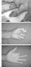
A young girl with loss of radial digits on the left hand and the ulnar digits on the right hand as a result of congenital ring constriction syndrome. The figure shows effective chuck pinch in both the hands irrespective of position of digits relative to the thumb.
Fig. 2
Fig. 2
This 60-year-old patient suffered a severe crush injury to the index finger and the middle finger. It was a zone 2 amputation of the index at the proximal interphalangeal joint. The middle finger was devascularized with a volar soft tissue defect, with intact interphalangeal joints and no fractures.
Fig. 3
Fig. 3
(A) The index finger was sacrificed and a neurovascular island flap was fashioned based on the ulnar digital artery and nerve of the index finger simultaneously providing soft tissue cover, revascularization and reinnervation to the middle finger without violation of any other donor site. (B,C) Long-term functional outcome following the procedure.
Fig. 4
Fig. 4
(A,B): Roller injury to the right hand. The distal phalanx of the thumb is amputated, the index finger is devascularized with soft tissue degloving. The middle finger had segmental injury with proximal phalanx fracture and devascularization; the ring finger had a proximal phalanx fracture. There was a dorsal skin defect over the hand. (C) The most severely injured finger (the index finger in this case) was harvested as the source of spare parts and divided into two halves. (D) Distal half of the index finger is microsurgically transferred to the thumb. Proximal index finger used as source of artery and nerve for grafting for revascularization and digital nerve reconstruction of the middle finger. Remnant skin from the index finger was used as a fillet flap to cover the dorsal skin defect. (E) A posterior interosseous artery flap performed at a second stage to cover the dorsal skin defect resulting from necrosis of the fillet flap harvested from the index finger. (F) Good long-term functional recovery following the procedure.
Fig. 5
Fig. 5
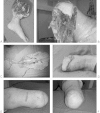
(A,B) A traumatic below-knee amputation with severe soft tissue crush injury to the proximal leg and intact knee joint. (C) An innervated medial plantar flap is harvested from the sole as a free flap. (D) The medial plantar flap transferred to reconstruct a below-knee amputation stump preserving the joint. (E,F) Long-term outcome showing a well-healed below-knee stump.
Fig. 6
Fig. 6
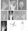
(A) Amputations of the index, middle, and ring fingers through the proximal interphalangeal joints. (B–D) The ring finger stump was harvested with both neurovascular pedicles to match the recipient site. It was split longitudinally to provide lengthening to the index and middle finger stump. The index and middle finger were initially syndactylized to preserve venous drainage and provide adequate skin cover. (E) X-ray of the hand following transfer of the ring finger stump to the index and the middle finger stumps with syndactylization of the fingers. (F) The fingers were separated at a second stage, with additional skin grafting resulting in a three-fingered hand. (G) X-ray of the hand after separation of the index and the middle fingers. (H) Schematic diagram of the fillet harvesting procedure. (I) Schematic diagram of transplantation of the filleted finger to the recipient index and middle finger stump and digit, respectively.

References

    1. Midgley R D, Entin M A. Management of mutilating injuries of the hand. Clin Plast Surg. 1976;3(1):99–109. - PubMed
    1. Büchler U. Traumatic soft-tissue defects of the extremities. Implications and treatment guidelines. Arch Orthop Trauma Surg. 1990;109(6):321–329. - PubMed
    1. Buchler U, Hastings H. New York, NY: Churchill Livingstone; 1993. Combined injuries; pp. 1631–1650.
    1. Soucacos P N. Indications and selection for digital amputation and replantation. J Hand Surg [Br] 2001;26(6):572–581. - PubMed
    1. Soucacos P N, Beris A E, Malizos K N, Vlastou C, Soucacos P K, Georgoulis A D. Transpositional microsurgery in multiple digital amputations. Microsurgery. 1994;15(7):469–473. - PubMed