. 2014 Oct;154(2):178-87.
doi: 10.1016/j.clim.2014.05.007. Epub 2014 May 27.

The role of antigen specificity in the binding of murine monoclonal anti-DNA antibodies to microparticles from apoptotic cells

Affiliations

The role of antigen specificity in the binding of murine monoclonal anti-DNA antibodies to microparticles from apoptotic cells

Anirudh J Ullal et al. Clin Immunol. 2014 Oct.

Abstract

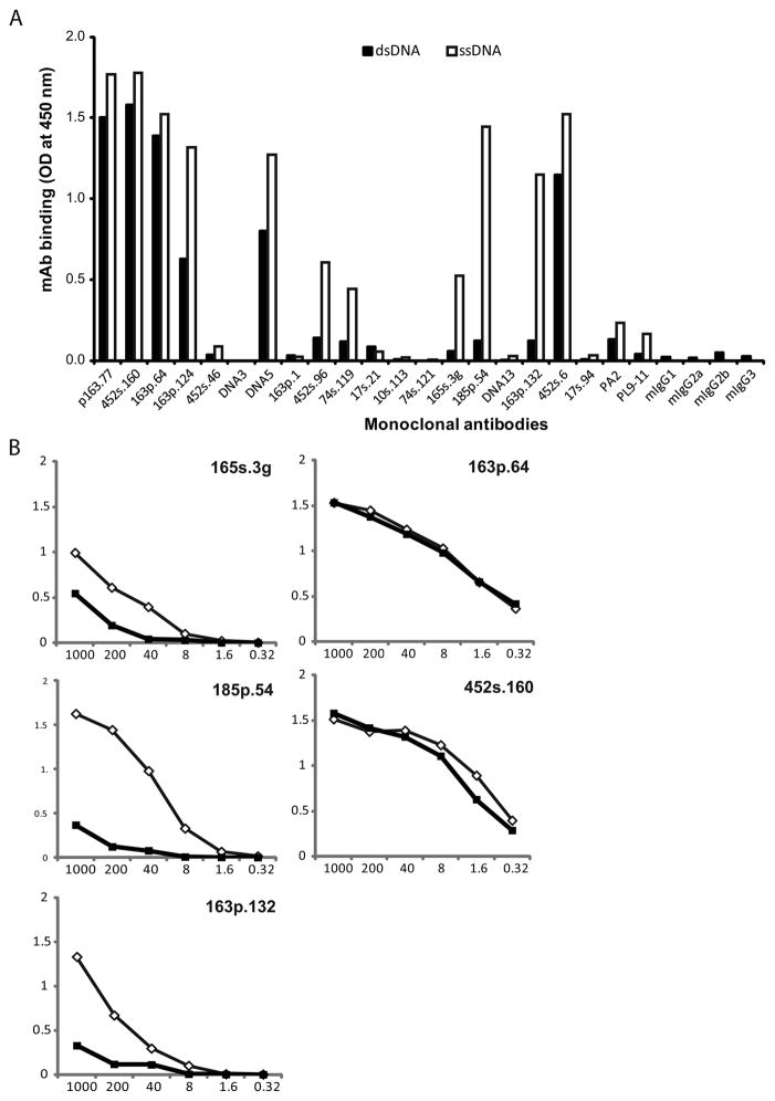
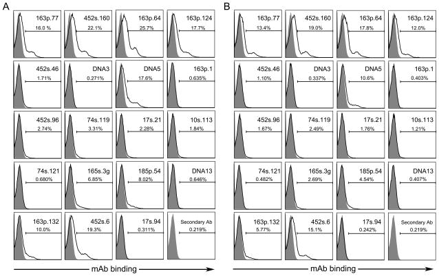
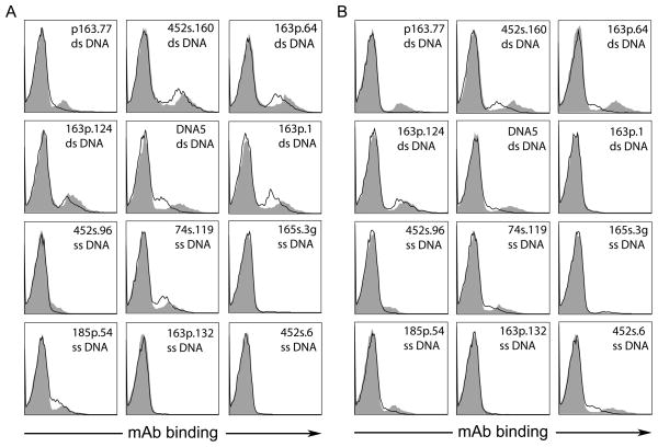
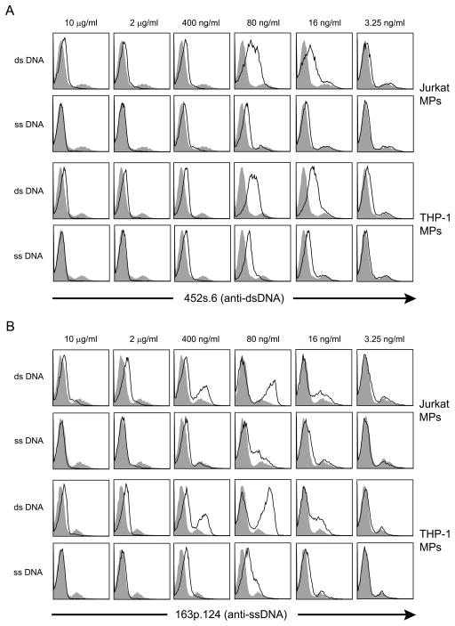
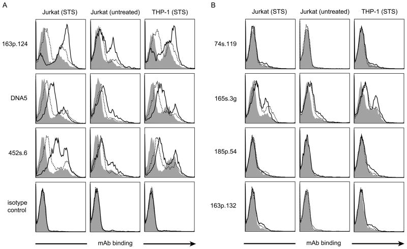
Antibodies to DNA (anti-DNA) are the serological hallmark of systemic lupus erythematosus and markers of underlying immune system disturbances. These antibodies bind to both single-stranded and double-stranded DNA, mediating pathogenesis by forming immune complexes. As shown recently, DNA in blood exists in both free and particulate forms, with DNA representing an important component of microparticles. Microparticles are membrane-bound vesicles containing nuclear molecules, released by membrane blebbing during cell death and activation. A panel of monoclonal NZB/NZW F1 anti-DNA antibodies was tested for binding to microparticles generated from apoptotic THP-1 and Jurkat cells. These studies showed that only certain anti-DNA antibodies in the panel, specific for double-stranded DNA, bound to microparticles. Binding to particles was reduced by soluble DNA or DNase treatment. Together, these results indicate that particle binding is a feature of only certain anti-DNA antibodies, reflecting immunochemical properties of the antibodies and the nature of the exposed DNA antigens.

Keywords: Anti-DNA;; Apoptosis; Autoimmunity;; Lupus;; Microvesicle;.


Figures

Figure 1
Figure 1
DNA specificity of monoclonal antibodies. A panel of 19 monoclonals (mAb) previously prepared from NZB/NZW F1 lupus mice was assayed by ELISA for binding specificity to ds or ssDNA. Microtiter plates were coated with 5 μg/ml of native (dsDNA) or denatured (ssDNA) calf-thymus DNA, treated with 250 ng of mAb (panel A) or five-fold serial dilutions (1000 – 0.32 ng) (panel B) in 100 μl of reaction volume and incubated for 1 h at 37°C. Peroxidase-labeled sheep anti-mouse IgG was used to measure bound antibodies. Black bars (A) or squares (B) represent mAb binding to dsDNA and white bars (A) or diamonds (B) show binding to ssDNA. PA2 and PL9-11 are control mAbs. Results are representative of three replicate experiments.
Figure 2
Figure 2
Binding of mAbs to MPs generated in vitro. Jurkat cells were treated with 1 μM staurosporine (A) or 10 μM etoposide (B) for 18 h to induce apoptosis. MPs were isolated from culture media by centrifugation and diluted in PBS to a concentration of 5000 MPs/μl for treatment with 500 ng of mAb for 1 h at 37°C. Binding of mAbs to MPs (black line) was detected by flow cytometry using R-phycoerythrin labeled goat anti-mouse IgG and compared to isotype controls (filled grey peaks). Presented data are representative of three replicate experiments.
Figure 3
Figure 3
The effects of nuclease treatment on the binding of mAbs to MPs. MPs prepared from Jurkat (A) and THP-1 (B) cells were treated with 100 U/ml of DNase (black line) or left untreated (filled peak). MPs were incubated with the mAbs (5 μg/ml) for 1 h at RT followed by staining with R-PE labeled goat anti-mouse IgG. Stained MPs were assayed by flow cytometry. Results are representative of four replicate experiments.
Figure 4
Figure 4
The effects of DNA pre-incubation on the binding of mAbs to MPs. At a concentration of 5μg/ml, two anti-DNA mAbs, 452s.6 (A) and 163p.124 (B), were incubated in five-fold serial dilutions of ssDNA or dsDNA (10 μg/ml – 3.25 ng/ml). The mAb was then added to a suspension of 500,000 MPs from THP-1 or Jurkat cells treated with staurosporine. MAb binding to MPs (black line) was detected by flow cytometry and compared to the binding of mAb with control pre-incubation (filled peak). Results are representative of experiments performed three times.
Figure 5
Figure 5
The effect of incubation of particles with DNA on the binding of mAb. Jurkat and THP-1 MPs from cultures treated with staurosporine were incubated in 10 μg/ml solutions of ssDNA (broken line), dsDNA (solid line) or left untreated (filled peak). Treated MPs were washed by centrifugation and resuspended in fresh PBS. MPs were incubated with 5 μg/ml of anti-dsDNA mAb (163p.124, DNA5 and 452s.6) (panel A) and anti-ssDNA mAb (74s.119, 165s.3g, 185p.54 and 163p.132) (panel B). Bound anti-DNA was detected by flow cytometry using R-PE labeled goat anti-mouse IgG. Results are representative of 3 experiments.

Similar articles

Cited by

References

    1. Hahn BH. Antibodies to DNA. N Engl J Med. 1998;338:1359–1368. - PubMed
    1. Tsokos GC. Systemic lupus erythematosus. N Engl J Med. 2011;365:2110–2121. - PubMed
    1. Jang YJ, Stollar BD. Anti-DNA antibodies: aspects of structure and pathogenicity. Cell Mol Life Sci. 2003;60:309–320. - PMC - PubMed
    1. Mortensen ES, Rekvig OP. Nephritogenic potential of anti-DNA antibodies against necrotic nucleosomes. J Am Soc Nephrol. 2009;20:696–704. - PubMed
    1. van der Vlag J, Berden JH. Lupus nephritis: role of antinucleosome autoantibodies. Semin Nephrol. 2011;31:376–389. - PubMed

Publication types