

A multifactorial role for P. falciparum malaria in endemic Burkitt's lymphoma pathogenesis
- PMID: 24874410
- PMCID: PMC4038605
- DOI: 10.1371/journal.ppat.1004170
A multifactorial role for P. falciparum malaria in endemic Burkitt's lymphoma pathogenesis
Abstract
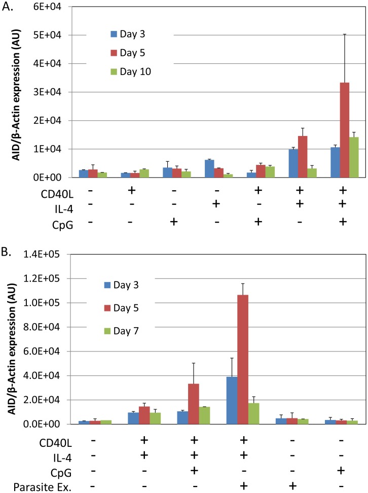
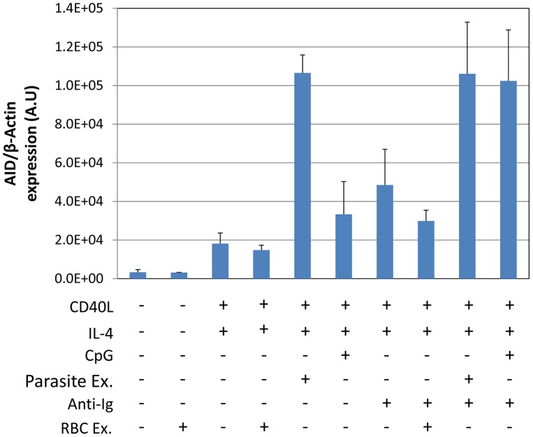
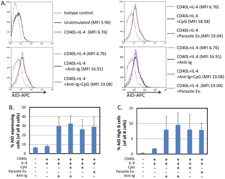
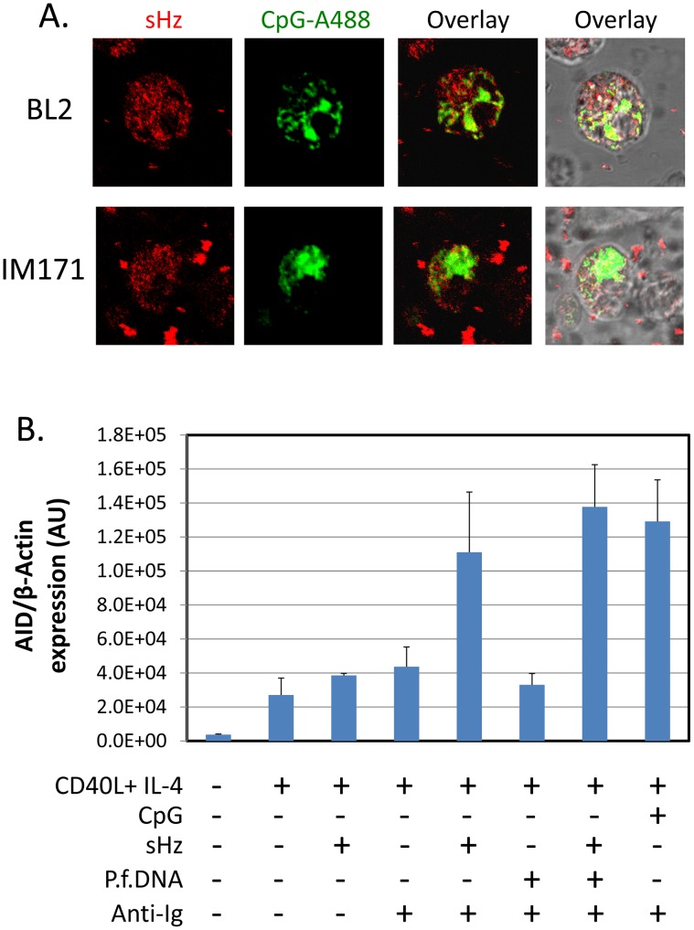
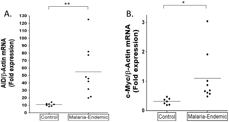
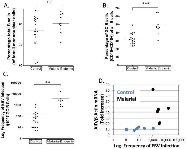
Endemic Burkitt's lymphoma (eBL) arises from the germinal center (GC). It is a common tumor of young children in tropical Africa and its occurrence is closely linked geographically with the incidence of P. falciparum malaria. This association was noted more than 50 years ago. Since then we have learned that eBL contains the oncogenic herpes virus Epstein-Barr virus (EBV) and a defining translocation that activates the c-myc oncogene. However the link to malaria has never been explained. Here we provide evidence for a mechanism arising in the GC to explain this association. Accumulated evidence suggests that eBL arises in the GC when deregulated expression of AID (Activation-induced cytidine deaminase) causes a c-myc translocation in a cell latently infected with Epstein-Barr virus (EBV). Here we show that P. falciparum targets GC B cells via multiple pathways to increase the risk of eBL. 1. It causes deregulated expression of AID, thereby increasing the risk of a c-myc translocation. 2. It increases the number of B cells transiting the GC. 3. It dramatically increases the frequency of these cells that are infected with EBV and therefore protected from c-myc induced apoptosis. We propose that these activities combine synergistically to dramatically increase the incidence of eBL in individuals infected with malaria.
Conflict of interest statement
The authors have declared that no competing interests exist.
Figures
Comment in
-
Parasite biology: Piecing it together.Nat Rev Microbiol. 2014 Jul;12(7):462-3. doi: 10.1038/nrmicro3303. Nat Rev Microbiol. 2014. PMID: 25068157 No abstract available.
Similar articles
-
Endemic Burkitt's lymphoma as a polymicrobial disease: new insights on the interaction between Plasmodium falciparum and Epstein-Barr virus.Semin Cancer Biol. 2009 Dec;19(6):411-20. doi: 10.1016/j.semcancer.2009.10.002. Epub 2009 Nov 6. Semin Cancer Biol. 2009. PMID: 19897039 Review.
-
Malaria, Epstein-Barr virus infection and the pathogenesis of Burkitt's lymphoma.Int J Cancer. 2017 Nov 1;141(9):1849-1855. doi: 10.1002/ijc.30885. Epub 2017 Jul 24. Int J Cancer. 2017. PMID: 28707393
-
The company malaria keeps: how co-infection with Epstein-Barr virus leads to endemic Burkitt lymphoma.Curr Opin Infect Dis. 2011 Oct;24(5):435-41. doi: 10.1097/QCO.0b013e328349ac4f. Curr Opin Infect Dis. 2011. PMID: 21885920 Free PMC article. Review.
-
Epstein-Barr virus and malaria upregulate AID and APOBEC3 enzymes, but only AID seems to play a major mutagenic role in Burkitt lymphoma.Eur J Immunol. 2022 Aug;52(8):1273-1284. doi: 10.1002/eji.202249820. Epub 2022 May 12. Eur J Immunol. 2022. PMID: 35503749 Free PMC article.
-
Endemic Burkitt's lymphoma: a polymicrobial disease?Nat Rev Microbiol. 2005 Feb;3(2):182-7. doi: 10.1038/nrmicro1089. Nat Rev Microbiol. 2005. PMID: 15685227 Review.
Cited by
-
Parasite-microbe-host interactions and cancer risk.PLoS Pathog. 2019 Aug 15;15(8):e1007912. doi: 10.1371/journal.ppat.1007912. eCollection 2019 Aug. PLoS Pathog. 2019. PMID: 31415672 Free PMC article. No abstract available.
-
Estimating the global burden of Epstein-Barr virus-related cancers.J Cancer Res Clin Oncol. 2022 Jan;148(1):31-46. doi: 10.1007/s00432-021-03824-y. Epub 2021 Oct 27. J Cancer Res Clin Oncol. 2022. PMID: 34705104 Free PMC article. Review.
-
Herpesviruses: Harmonious Pathogens but Relevant Cofactors in Other Diseases?Front Cell Infect Microbiol. 2018 May 25;8:177. doi: 10.3389/fcimb.2018.00177. eCollection 2018. Front Cell Infect Microbiol. 2018. PMID: 29888215 Free PMC article. Review.
-
EBV Persistence--Introducing the Virus.Curr Top Microbiol Immunol. 2015;390(Pt 1):151-209. doi: 10.1007/978-3-319-22822-8_8. Curr Top Microbiol Immunol. 2015. PMID: 26424647 Free PMC article. Review.
-
Comparison of Epstein-Barr virus and Kaposi's sarcoma-associated herpesvirus viral load in peripheral blood mononuclear cells and oral fluids of HIV-negative individuals aged 3-89 years from Uganda.Infect Agent Cancer. 2023 Jun 14;18(1):38. doi: 10.1186/s13027-023-00516-9. Infect Agent Cancer. 2023. PMID: 37316814 Free PMC article.
References
-
- Goldstein JA, Bernstein RL (1990) Burkitt's lymphoma and the role of Epstein-Barr virus. J Trop Pediatr 36: 114–120. - PubMed
-
- Klein U, Dalla-Favera R (2008) Germinal centres: role in B-cell physiology and malignancy. Nat Rev Immunol 8: 22–33. - PubMed
-
- Klein G (1983) Specific chromosomal translocations and the genesis of B-cell-derived tumors in mice and men. Cell 32: 311–315. - PubMed
-
- Leder P (1985) Translocations among antibody genes in human cancer. In: Lenoir GM, O'Conor GT, Olweny CLM, editors. Burkitt's Lymphoma: a human cancer model. New York: Oxford University Press. pp. 341–371.
MeSH terms
Grants and funding
LinkOut - more resources
Full Text Sources
Other Literature Sources
Miscellaneous
