

Detailed analysis of temporal features on contrast enhanced ultrasound may help differentiate intrahepatic cholangiocarcinoma from hepatocellular carcinoma in cirrhosis
- PMID: 24874413
- PMCID: PMC4038593
- DOI: 10.1371/journal.pone.0098612
Detailed analysis of temporal features on contrast enhanced ultrasound may help differentiate intrahepatic cholangiocarcinoma from hepatocellular carcinoma in cirrhosis
Abstract
Aim: To verify if detailed analysis of temporal enhancement patterns on contrast enhanced ultrasound (CEUS) may help differentiate intrahepatic cholangiocarcinoma (ICC) from hepatocellular carcinoma (HCC) in cirrhosis.
Methods: Thirty three ICC and fifty HCC in cirrhosis were enrolled in this study. The contrast kinetics of ICC and HCC was analyzed and compared.
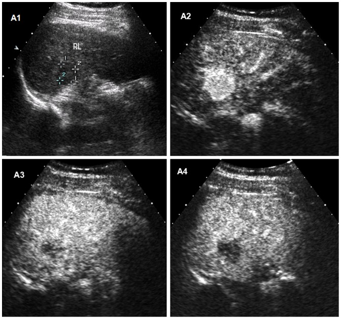
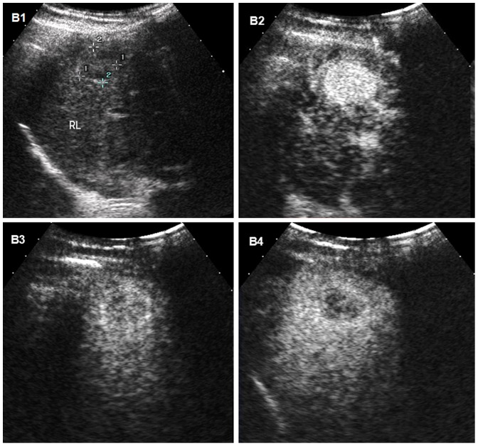
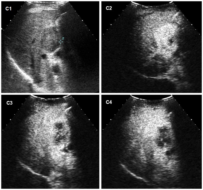
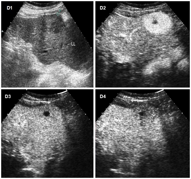
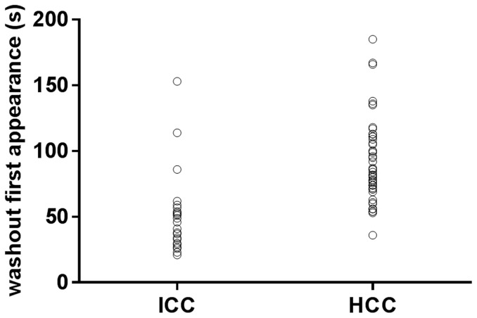
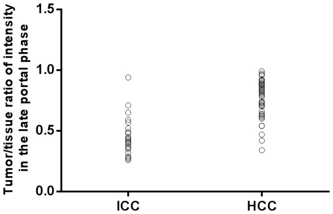
Results: Statistical analysis did not reveal significant difference between ICC and HCC in the time of contrast first appearance and arterial peak maximum time. ICC displayed much earlier washout than that of HCC (47.93±26.45 seconds vs 90.86±31.26 seconds) in the portal phase, and most ICC (87.9%) showed washout before 60 seconds than HCC (16.0%). Much more ICC (78.8%) revealed marked washout than HCC (12.0%) while most HCC (88.0%) showed mild washout or no washout in late part of the portal phase (90-120 seconds). Twenty six out of thirty three ICC (78.8%) demonstrated both early washout(<60 seconds) and marked washout in late part of the portal phase, whereas, only six of fifty HCC (12.0%)showed these temporal enhancement features (p = 0.000).When both early washout and marked washout in the portal phase are taken as diagnostic criterion for ICC, the diagnostic sensitivity, specificity, positive predictive value, negative predictive value and accuracy were 78.8%,88.0%,81.3%,86.3%,and 84.3% respectively by CEUS.
Conclusions: Analysis of detailed temporal enhancement features on CEUS is helpful differentiate ICC from HCC in cirrhosis.If a nodule in cirrhotic liver displays hyper-enhancement in the arterial phase followed by early and marked washout in the portal phase, the nodule is highly suspicious of ICC rather than HCC.
Conflict of interest statement
Figures
Similar articles
-
Applications of Dynamic Contrast-Enhanced Ultrasound in Differential Diagnosis of Hepatocellular Carcinoma and Intrahepatic Cholangiocarcinoma in Non-cirrhotic Liver.Ultrasound Med Biol. 2023 Aug;49(8):1780-1788. doi: 10.1016/j.ultrasmedbio.2023.03.026. Epub 2023 May 6. Ultrasound Med Biol. 2023. PMID: 37156676
-
CEUS in hepatocellular carcinoma and intrahepatic cholangiocellular carcinoma in 320 patients - early or late washout matters: a subanalysis of the DEGUM multicenter trial.Ultraschall Med. 2015 Apr;36(2):132-9. doi: 10.1055/s-0034-1399147. Epub 2015 Mar 26. Ultraschall Med. 2015. PMID: 25812115
-
Differentiation of intrahepatic cholangiocarcinoma from hepatocellular carcinoma in high-risk patients: A predictive model using contrast-enhanced ultrasound.World J Gastroenterol. 2018 Sep 7;24(33):3786-3798. doi: 10.3748/wjg.v24.i33.3786. World J Gastroenterol. 2018. PMID: 30197484 Free PMC article.
-
Differentiation between hepatocellular carcinoma and intrahepatic cholangiocarcinoma using contrast-enhanced ultrasound: A systematic review and meta-analysis.Clin Hemorheol Microcirc. 2021;79(2):293-309. doi: 10.3233/CH-211145. Clin Hemorheol Microcirc. 2021. PMID: 33935070
-
Contrast-enhanced ultrasound in the diagnosis of nodules in liver cirrhosis.World J Gastroenterol. 2014 Apr 7;20(13):3590-6. doi: 10.3748/wjg.v20.i13.3590. World J Gastroenterol. 2014. PMID: 24707142 Free PMC article. Review.
Cited by
-
Contrast-enhanced ultrasound imaging features and clinical characteristics of combined hepatocellular cholangiocarcinoma: comparison with hepatocellular carcinoma and cholangiocarcinoma.Ultrasonography. 2020 Oct;39(4):356-366. doi: 10.14366/usg.19093. Epub 2020 Mar 13. Ultrasonography. 2020. PMID: 32407611 Free PMC article.
-
Contrast-enhanced ultrasonography: advance and current status in abdominal imaging.Ultrasonography. 2015 Jan;34(1):3-18. doi: 10.14366/usg.14034. Epub 2014 Sep 12. Ultrasonography. 2015. PMID: 25342120 Free PMC article. Review.
-
Contrast-enhanced Ultrasound Features of Intrahepatic Cholangiocarcinoma: A New Perspective.Sci Rep. 2019 Dec 18;9(1):19363. doi: 10.1038/s41598-019-55857-6. Sci Rep. 2019. PMID: 31852947 Free PMC article.
-
Imaging Diagnosis of Hepatocellular Carcinoma: Recent Advances of Contrast-Enhanced Ultrasonography with SonoVue®.Liver Cancer. 2016 Feb;5(1):55-66. doi: 10.1159/000367748. Epub 2015 Dec 18. Liver Cancer. 2016. PMID: 29234627 Free PMC article. Review.
-
Diagnosis of Hepatic Angiomyolipoma by Combination of Baseline and Contrast-Enhanced Ultrasound--A Prospective Study in Non-Cirrhotic Patients.PLoS One. 2015 Jul 6;10(7):e0132290. doi: 10.1371/journal.pone.0132290. eCollection 2015. PLoS One. 2015. PMID: 26147859 Free PMC article.
References
-
- Rimola J, Forner A, Tremosini S, Reig M, Vilana R, et al. (2012) Non-invasive diagnosis of hepatocellular carcinoma 2 cm in cirrhosis. Diagnostic accuracy assessing fat, capsule and signal intensity at dynamic MRI. J Hepatol 56: 1317–1323. - PubMed
-
- Serste T, Barrau V, Ozenne V, Vullierme MP, Bedossa P, et al. (2012) Accuracy and disagreement of computed tomography and magnetic resonance imaging for the diagnosis of small hepatocellular carcinoma and dysplastic nodules: role of biopsy. Hepatology 55: 800–806. - PubMed
-
- Shaib Y, El-Serag HB (2004) The epidemiology of cholangiocarcinoma. Semin Liver Dis 24: 115–125. - PubMed
-
- Bruix J, Sherman M (2005) Management of hepatocellular carcinoma. Hepatology 42: 1208–1236. - PubMed
-
- EASL-EORTC (2012) Clinical practice guidelines Management of hepatocellular carcinoma. J Hepatol 56: 908–943. - PubMed
MeSH terms
Substances
LinkOut - more resources
Full Text Sources
Other Literature Sources
Medical
