

doi: 10.3174/ajnr.A3987.
Epub 2014 May 29.
Interhypothalamic adhesion: a series of 13 cases
Affiliations
- PMID: 24874532
- PMCID: PMC7966237
- DOI: 10.3174/ajnr.A3987
Item in Clipboard
Interhypothalamic adhesion: a series of 13 cases
AJNR Am J Neuroradiol.
2014 Oct.
Abstract
Interhypothalamic adhesion is a newly described disease entity, characterized by an abnormal parenchymal band connecting the medial margins of the hypothalami across the third ventricle. Additional anomalies, including cleft palate, gray matter heterotopia, cerebellar hypoplasia, optic atrophy, hippocampal under-rotation, and white matter lesions, may coexist. The purpose of this clinical report is to describe the imaging findings from a series of 13 patients with interhypothalamic adhesions discovered on brain MR imaging.
© 2014 by American Journal of Neuroradiology.
Figures
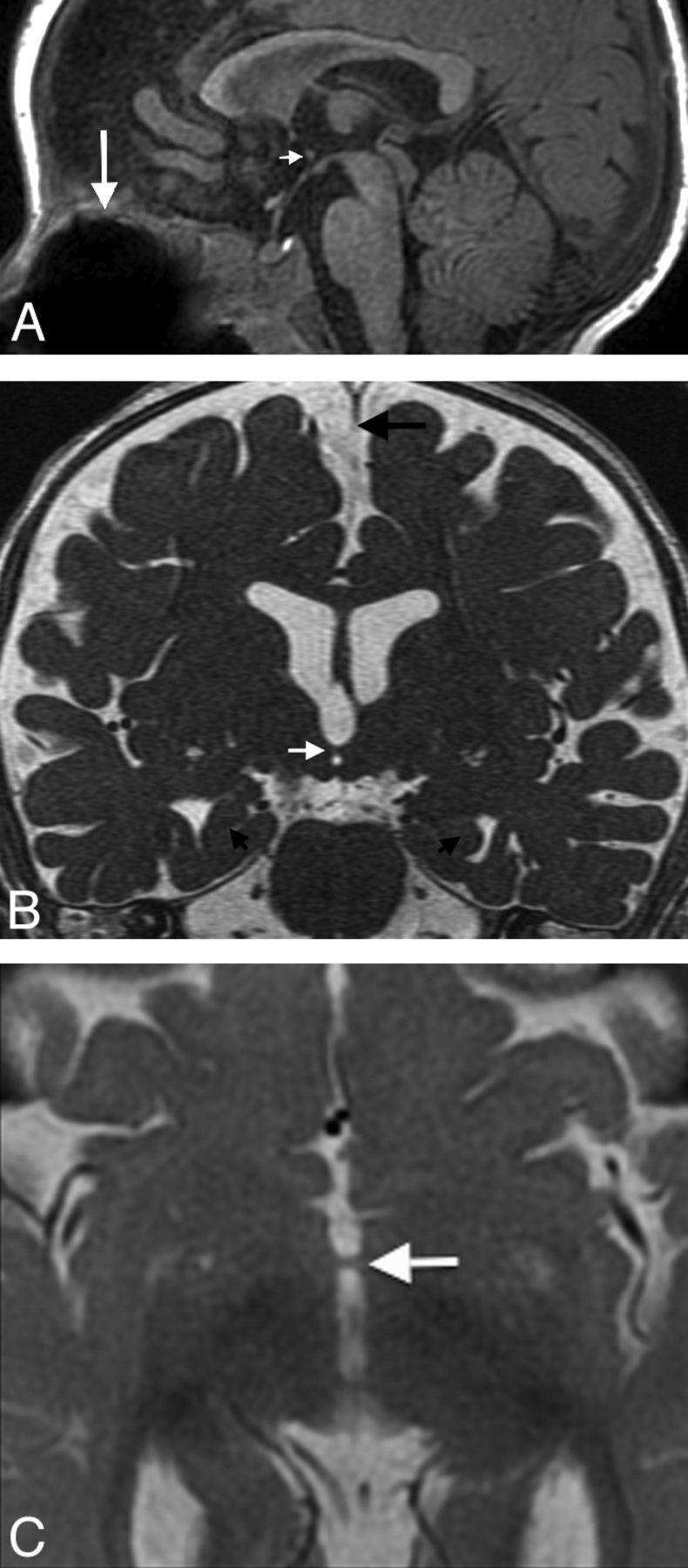
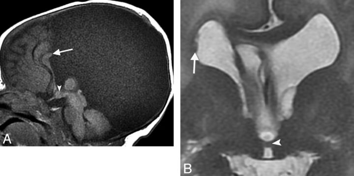
A, Sagittal fast-spoiled gradient recalled brain volume T1WI (TR/TE/TI, 10/4/450 ms) showing a parenchymal band representing an interhypothalamic adhesion in the third ventricle between the anterior commissure and mammillary body (small arrow). The pituitary infundibulum, adenohypophysis, and neurohypophysis are normal. Note metallic susceptibility artifacts in the roof of the oral cavity from prior cleft palate repair (large arrow). B, Coronal FIESTA with phase cycling (TR/TE, 10/4 ms) demonstrates an IHA traversing the third ventricle (white arrow), oblique axes of the hippocampal heads representing under-rotation (small black arrows), and incomplete formation of the falx cerebri (large black arrow). C, Axial T2WI (TR/TE, 5000/85 ms) shows an IHA connecting the medial hypothalamic margins across the third ventricle (arrow).
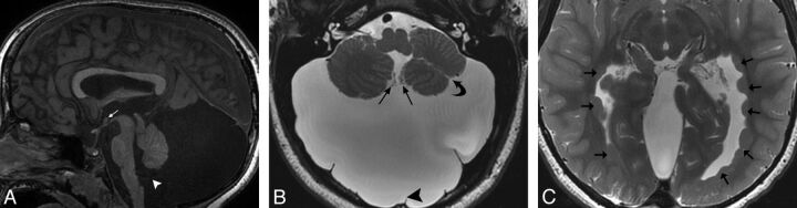
A, Sagittal SPGR T1WI (TR/TE/IR, 8/3/450 ms) demonstrating a nodular structure isointense to gray matter consistent with an IHA (white arrow) located at the level of the upper midbrain in the anterior/inferior third ventricle, spanning to the third ventricular floor. A Blake pouch cyst is present, enlarging the posterior fossa and communicating with the fourth ventricle via a widened foramen of Magendie (white arrowhead) and without accompanying vermian malrotation. Note also generalized vermian and brain stem hypoplasia. B, Axial T2 fast-spoiled gradient recalled image (TR/TE, 3049/99 ms) shows a large retrocerebellar cystic lesion markedly enlarging the posterior fossa, associated with an incomplete falx cerebelli (arrowhead). Choroid plexus is seen extending from the foramen of Magendie along its anteromedial walls (straight arrows), consistent with a Blake pouch cyst. The cerebellum is dysplastic with irregular foliation and a left cerebellar hemispheric cleft (curved arrow). C, Axial T2 fast-spoiled gradient recalled scan (TR/TE, 3049/99 ms) depicts periventricular nodular heterotopia along the lateral ventricular atria and occipital and temporal horns (arrows).
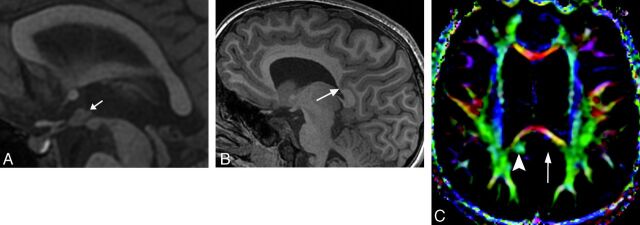
A, Sagittal SPGR T1WI (TR/TE/IR, 8/3/450 ms) demonstrating a nodular structure isointense to gray matter consistent with IHA (arrow) located at the level of the upper midbrain in the inferior third ventricle, extending to the tuber cinereum. B, Sagittal SPGR T1WI (TR/TE/IR, 8/3/450 ms) demonstrates an unusual deep parietal sulcus extending through the cingulate gyrus and distorting the callosal splenium architecture (arrow). C, Axial directionally encoded color map of DTI data (15 directions of encoding; TR/TE, 10,000/82 ms) shows absence of the normal green hue of the left cingulum isthmus (long arrow). Note the normal right cingulum isthmus (arrowhead).
A, Sagittal SPGR T1WI (TR/TE/IR, 11/5/500 ms) demonstrating a small nodular structure representing an IHA located in the midanterior third ventricle in the expected area of the paraventricular nucleus (arrowhead). Parenchymal stigmata of a Chiari II malformation include a small posterior fossa, tectal beaking (curved arrow), and mild corpus callosum dysgenesis. In addition, partial rhombencephalosynapsis is present with loss of the normal architecture of the posterior vermis (straight arrow). B, Coronal fat-saturated T2WI (TR/TE, 3931/100 ms) shows continuous transverse cerebellar hemispheric folia and fissures extending across the midline, representing absence of the posterior vermis and partial rhombencephalosynapsis.
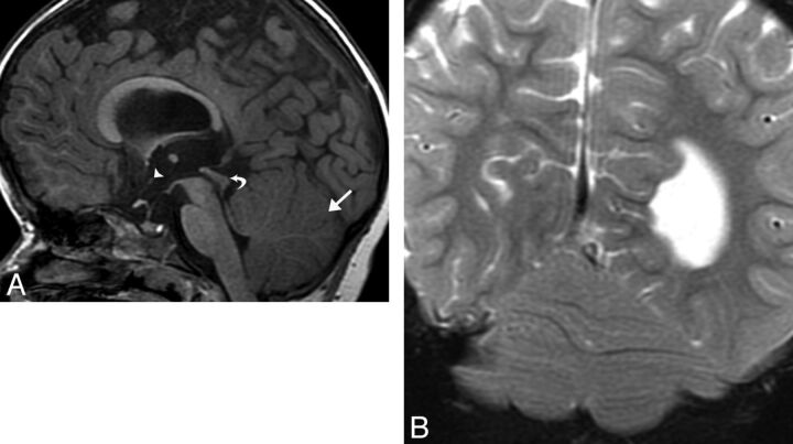
A, Sagittal T1WI (TR/TE, 400/9 ms) depicting a nodular structure occupying the anterior/inferior third ventricle, consistent with an IHA (arrowhead). A large interhemispheric meningeal cyst is associated with corpus callosal dysgenesis (arrow). B, Coronal T2WI (TR/TE, 2217/103 ms) shows a horizontal parenchymal band adjoining the medial thalami, representing an IHA (arrowhead), and periventricular nodular heterotopia (arrow).
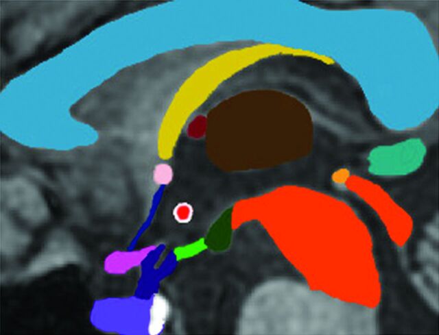
Midline sagittal graphic depicting the hypothalamus and neighboring structures. With the exception of the mammillary bodies (dark green) and tuber cinereum (bright green), the hypothalami are not visible at midline. The location of the IHA (bright red/white margin) is typical, centered in the anterior/inferior third ventricle. Choroid plexus is present in the anterior/superior third ventricle (dark red). The corpus callosum (light blue), fornix (yellow), thalamus (brown), midbrain (dark orange), posterior commissure (bright orange), pineal gland (teal), lamina terminalis (dark blue), anterior commissure (light pink), optic chiasm (dark pink), pituitary stalk (dark purple), adenohypophysis (light purple), and neurohypophysis (white) are represented.
References
-
- Lemaire J, Nezzar H, Sakka L, et al. . Maps of the adult human hypothalamus. Surg Neurol Int 2013;4(suppl 3):S156–63 - PubMed
-
- Nieuwenhuys R, Voogd J, van Huijzen C. The Human Central Nervous System. 4th ed. Berlin: Springer-Verlag; 2008:289–90
-
- Tortori-Donati P, Rossi A, Biancheri R. Brain malformations. In: Tortori-Donati P, Rossi A, eds. Pediatric Neuroradiology: Brain, Head, Neck and Spine. Berlin: Springer-Verlag; 2009:86–94
MeSH terms
LinkOut - more resources
Full Text Sources
Other Literature Sources
Medical