. 2014 Sep:69:192-9.
doi: 10.1016/j.nbd.2014.05.024. Epub 2014 May 27.

Anti-neutrophil antibody enhances the neuroprotective effects of G-CSF by decreasing number of neutrophils in hypoxic ischemic neonatal rat model

Affiliations

Anti-neutrophil antibody enhances the neuroprotective effects of G-CSF by decreasing number of neutrophils in hypoxic ischemic neonatal rat model

Desislava M Doycheva et al. Neurobiol Dis. 2014 Sep.

Abstract

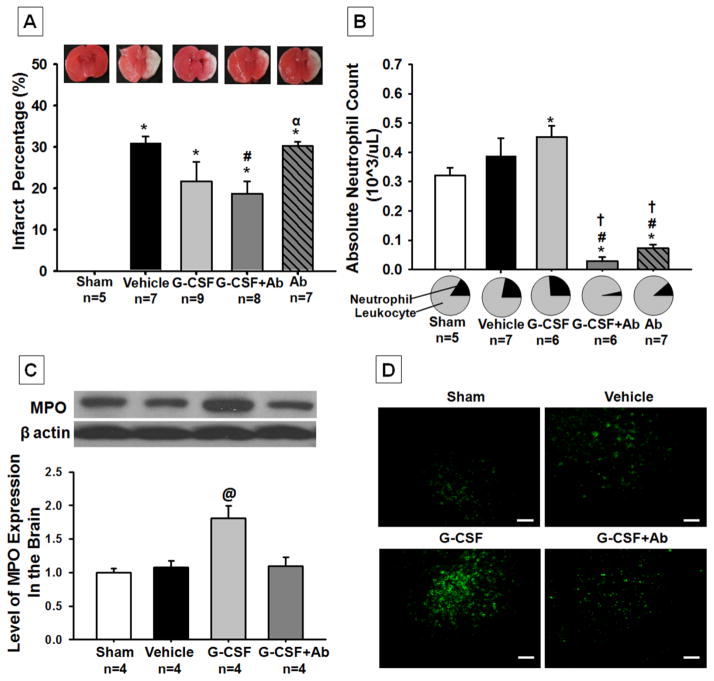
Objectives: Neonatal hypoxia ischemia (HI) is an injury that can lead to neurological impairments such as behavioral and learning disabilities. Granulocyte-colony stimulating factor (G-CSF) has been demonstrated to be neuroprotective in ischemic stroke however it has also been shown to induce neutrophilia, ultimately exacerbating neuronal injury. Our hypothesis is that coadministration of anti-neutrophil antibody (Ab) with G-CSF will decrease blood neutrophil counts thereby reducing infarct volume and improving neurological function post HI brain injury.

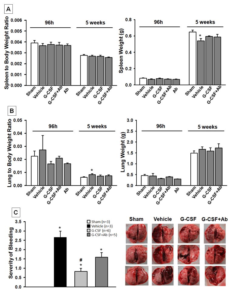
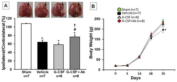
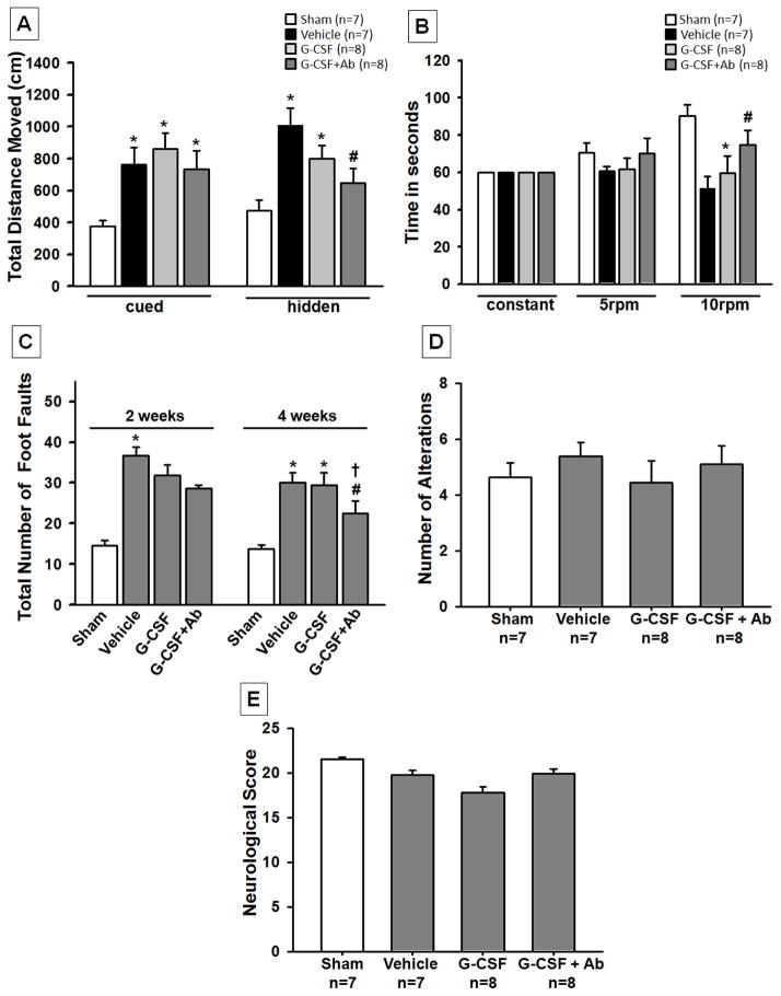
Methods: Rat pups were subjected to unilateral carotid artery ligation followed by 2.5h of hypoxia. Animals were randomly assigned to five groups: Sham (n=15), vehicle (HI, n=15), HI with G-CSF treatment (n=15), HI with G-CSF+Ab treatment (n=15), and HI with Ab treatment (n=15). Ab (325μg/kg) was administered intraperitoneally while G-CSF (50μg/kg) was administered subcutaneously 1h post HI followed by daily injections for 3 consecutive days. Animals were euthanized at 96h post HI for blood neutrophil counts and brain infarct volume measurements as well as at 5weeks for neurological function testing and brain weight measurements. Lung and spleen weights at both time points were further analyzed.

Results: The G-CSF treatment group showed tendencies to reduce infarct volume and improve neurological function while significantly increasing neutrophil counts. On the other hand, the G-CSF+Ab group significantly reduced infarct volume, improved neurological function and decreased neutrophil counts. The Ab alone group showed reversal of the neuroprotective effects of the G-CSF+Ab group. No significant differences were found in peripheral organ weights between groups.

Conclusion: Our data suggest that coadministration of G-CSF with Ab not only prevented brain atrophy but also significantly improved neurological function by decreasing blood neutrophil counts. Hence the neuroprotective effects of G-CSF may be further enhanced if neutrophilia is avoided.

Keywords: Anti-neutrophil antibody (Ab); Granulocyte-colony stimulating factor (G-CSF); Hypoxia–ischemia (HI); Neonatal; Neurological function; Neutrophil.


Conflict of interest statement

The authors declare they have no conflict of interest.

Figures

Fig. 1
Fig. 1. Effects of Treatment on Infarct Volume (A) Blood Neutrophil Counts (B) Brain Neutrophil Counts (C) and Representative Pictures of Infiltrating Neutrophils in Brain (D) at 96h post HI
Postnatal day-10 rats were subjected to HI. Intraperitoneal (IP) treatment with G-CSF, G-CSF+Ab or Ab began at 1h post HI and continued daily for 4 days. (A) While the G-CSF treatment group showed a tendency to reduce infarction, the G-CSF+Ab group significantly reduced infarct volume when compared to vehicle (p<0.004, 30.957±1.564 vehicle vs. 18.746±2.983 G-CSF+Ab). (B) Absolute neutrophil counts were significantly increased in the G-CSF treatment group when compared to vehicle and were successfully reduced in the combinational treatment (G-CSF+Ab) and Ab alone group (Data represent +/− SEM; *p<0.05 versus sham, #p<0.05 versus vehicle, †p<0.05 versus G-CSF, α p<0.05 versus G-CSF + Ab). (C) Western Blot analysis showed that MPO expression (marker for neutrophils) was significantly increased in the G-CSF treatment group when compared to all other groups (Data represent +/− SEM; @p<0.05 versus sham, vehicle and G-CSF+Ab). (D) Immunohistochemical staining of MPO (neutrophils) in the brain showed a significant increase in neutrophil counts after G-CSF treatment compared to other groups (White bar in picture represents 100μm).
Fig. 2
Fig. 2. Effects of Treatment on Organ Weight at 96h and 5 weeks post HI and Lung Bleeding
The absolute spleen weight in the vehicle group was significantly lower than the sham group at 5 weeks post HI. There was no intergroup difference in the spleen to body weight ratio at 96h or 5 weeks (A). There was a significant increase in the lung to body weight ratio in vehicle group at 5 weeks post HI when compared to sham (B). Treatment groups improved lung to body weight ratio (p>0.05 vs. Sham). The graph in part (C) shows the severity of lung bleeding at 5 weeks post HI. It is graded on a scale from 0 to 3, with zero meaning no bleeding at all and 3 meaning more than ¾ of lung bleeding. The vehicle group showed severe bleeding compared to sham; while G-CSF significantly reduced lung bleeding compared to vehicle, G-CSF+Ab treatment group also showed tendency to reduce bleeding but significance was not reached (Data represent +/− SEM; *p<0.05 vs. sham, #p<0.05 vs. vehicle).
Fig. 3
Fig. 3. Effects of Treatment on Brain Atrophy (A) and Body Weight (B) at 5 weeks post HI
(A) Significant loss of right-to-left hemispheric (RH: LH) weight ratio is evident in vehicle rats and significantly improved with G-CSF+Ab treatment at 5 weeks post HI. (Representative pictures shown; Data represent +/− SEM; *p<0.05 versus sham, #p<0.05 versus vehicle, †p<0.05 versus G-CSF). (B) At 5 weeks, the vehicle group showed a loss in body weight compared to sham and treatment groups. (Data represent +/− SEM; *p<0.05 vehicle vs. sham, G-CSF and G-CSF+Ab).
Fig. 4
Fig. 4. Functional Outcome post HI
The combinational treatment group (G-CSF+Ab) showed significant improvement in neurological function according to (A) Morris Watermaze test, where spatial memory and learning was evaluated at 5 weeks post HI, (B) Rota Rod where sensori-motor function was evaluated at 5 weeks and (C) Foot fault test, which shows the total number of foot faults at 4 weeks. The results show that ischemia-induced foot slips were significantly reduced by treatment with G-CSF+Ab. T-maze (D) and Modified Garcia Test (E) did not show any significant improvements among treatment groups although there was a tendency for improvement. (Data represent +/− SEM; *p<0.05 versus sham, #p<0.05 versus vehicle, †p<0.05 versus G-CSF).

References

    1. Barone FC, et al. Polymorphonuclear leukocyte infiltration into cerebral focal ischemic tissue: myeloperoxidase activity assay and histologic verification. J Neurosci Res. 1991;29(3):336–45. - PubMed
    1. Barth TM, Stanfield BB. The recovery of forelimb-placing behavior in rats with neonatal unilateral cortical damage involves the remaining hemisphere. J Neurosci. 1990;10(10):3449–59. - PMC - PubMed
    1. Beck H, et al. Participation of bone marrow-derived cells in long-term repair processes after experimental stroke. J Cereb Blood Flow Metab. 2003;23(6):709–17. - PubMed
    1. Bracewell M, Marlow N. Patterns of motor disability in very preterm children. Ment Retard Dev Disabil Res Rev. 2002;8(4):241–8. - PubMed
    1. Broudy VC, et al. Human umbilical vein endothelial cells display high-affinity c-kit receptors and produce a soluble form of the c-kit receptor. Blood. 1994;83(8):2145–52. - PubMed

Publication types

MeSH terms

Substances