. 2014 May 29;8(5):e2897.
doi: 10.1371/journal.pntd.0002897. eCollection 2014.

Scabies mites alter the skin microbiome and promote growth of opportunistic pathogens in a porcine model

Affiliations

Scabies mites alter the skin microbiome and promote growth of opportunistic pathogens in a porcine model

Pearl M Swe et al. PLoS Negl Trop Dis. .

Abstract

Background: The resident skin microbiota plays an important role in restricting pathogenic bacteria, thereby protecting the host. Scabies mites (Sarcoptes scabiei) are thought to promote bacterial infections by breaching the skin barrier and excreting molecules that inhibit host innate immune responses. Epidemiological studies in humans confirm increased incidence of impetigo, generally caused by Staphylococcus aureus and Streptococcus pyogenes, secondary to the epidermal infestation with the parasitic mite. It is therefore possible that mite infestation could alter the healthy skin microbiota making way for the opportunistic pathogens. A longitudinal study to test this hypothesis in humans is near impossible due to ethical reasons. In a porcine model we generated scabies infestations closely resembling the disease manifestation in humans and investigated the scabies associated changes in the skin microbiota over the course of a mite infestation.

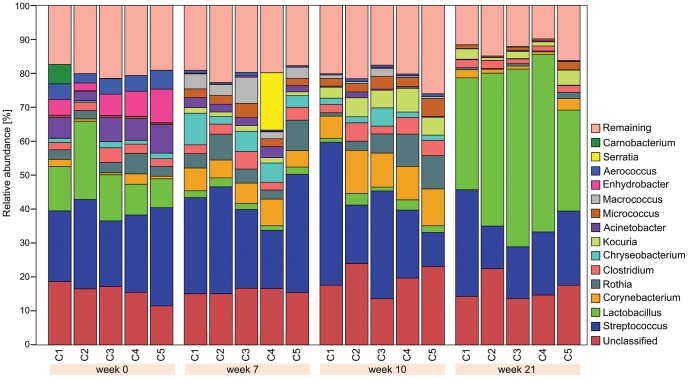
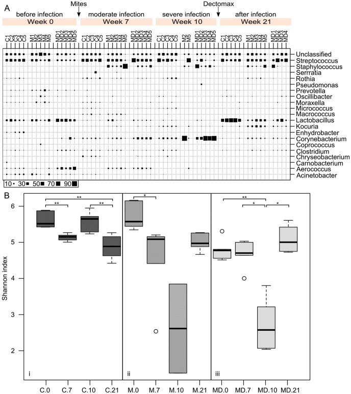
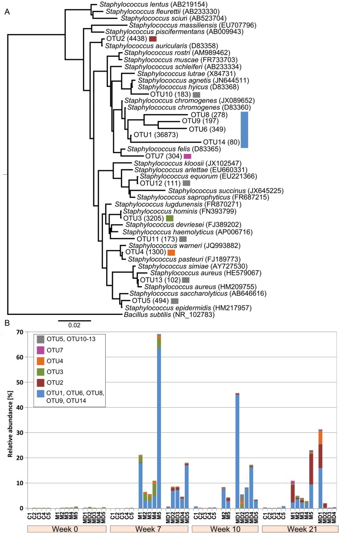
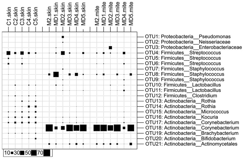
Methodology/principal findings: In a 21 week trial, skin scrapings were collected from pigs infected with S. scabies var. suis and scabies-free control animals. A total of 96 skin scrapings were collected before, during infection and after acaricide treatment, and analyzed by bacterial 16S rDNA tag-encoded FLX-titanium amplicon pyrosequencing. We found significant changes in the epidermal microbiota, in particular a dramatic increase in Staphylococcus correlating with the onset of mite infestation in animals challenged with scabies mites. This increase persisted beyond treatment from mite infection and healing of skin. Furthermore, the staphylococci population shifted from the commensal S. hominis on the healthy skin prior to scabies mite challenge to S. chromogenes, which is increasingly recognized as being pathogenic, coinciding with scabies infection in pigs. In contrast, all animals in the scabies-free cohort remained relatively free of Staphylococcus throughout the trial.

Conclusions/significance: This is the first experimental in vivo evidence supporting previous assumptions that establishment of pathogens follow scabies infection. Our findings provide an explanation for a biologically important aspect of the disease pathogenesis. The methods developed from this pig trial will serve as a guide to analyze human clinical samples. Studies building on this will offer implications for development of novel intervention strategies against the mites and the secondary infections.


Conflict of interest statement

The authors have declared that no competing interests exist.

Figures

Figure 1
Figure 1. Relative abundance of the most predominant bacterial genera in the pinna of porcine ears at various time points in the absence of mites.
At week 0, Streptococcus was the most dominant genus, followed by Lactobacillus, Actinectobacter, Enhydrobacter, Aerococcus and Rothia in decreasing abundance. At week 7, Streptococcus was still the most dominant genus, followed by smaller abundance of Corynebacterium, Chryseobacterium, Rothia, Clostridium and Lactobacillus. Similarly at week 10, Streptococcus was the dominant genus, followed by Corynebacterium, Rothia, Kocouria, Clostridium and Lactobacillus. At week 21, Lactobacillus was the most dominant, followed by Streptococcus and Kocuria. Only genera, with a relative abundance of ≥10% in at least one sample are illustrated.
Figure 2
Figure 2. (a) Composition of the microbial communities before, during and after scabies mite infection.
Microbial community structure was analysed by 454 pyrosequencing of the 16S rRNA gene of the total DNA, extracted from the skin scrapings of the pinna of pig ears. Samples were taken before the scabies infection from all cohorts at week 0. Cohorts M and MD were then challenged with an equal dosage of S. scabiei var. suis in both ears. Visible signs of scabies infection developed by week 7, which progressed to severe form of infection by week 10. Scabies infection was treated with an acaricide (Dectomax®) and skin was allowed to heal. Final skin scrapings were taken at week 21. Bubble volumes correlate with the abundance (%) of the each genus detected in the sample. (b) Effect of scabies mite infection on the microbial community diversity visualised by Shannon index for all samples from the cohorts C (i, mite free), M (ii, mite infested), MD (iii, mite infested and Dexamethasone treated). Community diversity is expressed as the mean Shannon index (OTU level). Paired t-test was carried out for pigs where both time points were available (M.10: n = 2, MD: n = 4, other: n = 5). Significant differences are annotated by *: p<0.05, **:p<0.01, ***: p<0.001.
Figure 3
Figure 3. (a) Phylogenetic tree of Staphylococcus indicating tentative species.
(b) Relative abundance of staphylococcal species in each sample. (a) A phylogenetic tree was constructed for OTUs assigned to the genus Staphylococcus using known reference 16S rRNA genes. The accession is noted in bracket for each reference species. The number in brackets following the OTU name indicates the read counts within the OTU. The tree was rooted with the outlier Bacillus subtilis. Several OTUs (1, 6, 8, 9, 14) are in close proximity to the 16S rRNA gene of S. chromogenes. (b) Bar chart demonstrating the relative abundance of each OTU in the pig cohorts over time. An extensive change of the OTUs is measured in the scabies infected cohort M and MD from week 7 onwards. The change was irreversible despite treatment. In particular, OTU1, OTU6, OTU8, OTU9, OTU14, all related to S. chromogenes, are dominating the skin microbiota.
Figure 4
Figure 4. (a) Phylogenetic tree of Streptococcus indicating tentative species.
(b) Relative abundance of streptococcal species in each sample. (a) A phylogenetic tree was constructed for OTUs assigned to the genus Streptococcus using known reference 16S rRNA genes. The accession is noted in bracket for each reference species. The number in brackets following the OTU name indicates the read counts within the OTU. The tree was rooted with the outlier Lactococcus lactis. (b) Bar chart demonstrating the relative abundance of each OTU in the pig cohorts over time. The graph depicts that the OTUs are overall stable between the three cohorts within each week and over time, irrespective of the presence or absence of mites.
Figure 5
Figure 5. Microbial composition of skin samples and isolated scabies mites collected from severely scabies infested crusted sites (week 10) and control animals.
Bubble plot of the taxonomic profile as calculated for the OTU level. Only OTUs with a relative abundance of ≥2% in at least one sample are shown. Each OTU is denoted with the taxonomic classification according to the RDP Classifier (confidence value ≥0.6). The taxonomic composition between the healthy skin and the mite containing skin as well as the washed mites is differing mainly based on the relative abundance of Corynebacteria.

References

    1. Engelman D, Kiang K, Chosidow O, McCarthy J, Fuller C, et al. (2013) Toward the Global Control of Human Scabies: Introducing the International Alliance for the Control of Scabies. PLoS Negl Trop Dis 7(8): e2167 doi:10.1371/journal.pntd.0002167 - DOI - PMC - PubMed
    1. Hay RJ, Steer AC, Engelman D, Walton S (2012) Scabies in the developing world—its prevalence, complications, and management. Clin Microbiol Infect 18: 313–323. - PubMed
    1. Fuller LC (2013) Epidemiology of scabies. Curr Opin Infect Dis 26: 123–126. - PubMed
    1. Hicks MI, Elston DM (2009) Scabies. Dermatol Ther 22: 279–292. - PubMed
    1. Makigami K, Ohtaki N, Ishii N, Yasumura S (2009) Risk factors of scabies in psychiatric and long-term care hospitals: a nationwide mail-in survey in Japan. J Dermatol 36: 491–498. - PubMed

Publication types

MeSH terms