Review
. 2015 Jan 7;10(1):135-46.
doi: 10.2215/CJN.05760513. Epub 2014 May 29.

Collecting duct principal cell transport processes and their regulation

Affiliations
Review

Collecting duct principal cell transport processes and their regulation

David Pearce et al. Clin J Am Soc Nephrol. .

Abstract

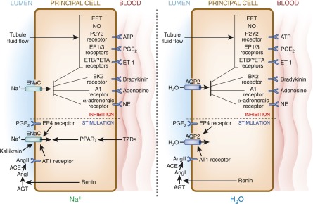
The principal cell of the kidney collecting duct is one of the most highly regulated epithelial cell types in vertebrates. The effects of hormonal, autocrine, and paracrine factors to regulate principal cell transport processes are central to the maintenance of fluid and electrolyte balance in the face of wide variations in food and water intake. In marked contrast with the epithelial cells lining the proximal tubule, the collecting duct is electrically tight, and ion and osmotic gradients can be very high. The central role of principal cells in salt and water transport is reflected by their defining transporters-the epithelial Na(+) channel (ENaC), the renal outer medullary K(+) channel, and the aquaporin 2 (AQP2) water channel. The coordinated regulation of ENaC by aldosterone, and AQP2 by arginine vasopressin (AVP) in principal cells is essential for the control of plasma Na(+) and K(+) concentrations, extracellular fluid volume, and BP. In addition to these essential hormones, additional neuronal, physical, and chemical factors influence Na(+), K(+), and water homeostasis. Notably, a variety of secreted paracrine and autocrine agents such as bradykinin, ATP, endothelin, nitric oxide, and prostaglandin E2 counterbalance and limit the natriferic effects of aldosterone and the water-retaining effects of AVP. Considerable recent progress has improved our understanding of the transporters, receptors, second messengers, and signaling events that mediate principal cell responses to changing environments in health and disease. This review primarily addresses the structure and function of the key transporters and the complex interplay of regulatory factors that modulate principal cell ion and water transport.

Keywords: collecting duct; epithelial cell; principal cell; renal physiology.


Figures

Figure 1.
Figure 1.
Hormonal regulation of ion transport in a typical principal cell. Principal cells respond to a variety of stimuli to control Na+ and K+ transport. Aldosterone has the most pronounced effect. It acts through the MR to increase expression of the serine-threonine kinase SGK1. Other hormones, including insulin, regulate SGK1 activity through the master kinase phosphatidylinositide 3-kinase (PI3K). SGK1 phosphorylates a variety of proteins; Nedd4-2 is shown, which is an ENaC inhibitor (see the text). SGK1 phosphorylation triggers interaction of Nedd4-2 with 14-3-3 proteins, and hence inhibition, by reducing its internalization and degradation. ENaC surface expression and activity are governed by a multiprotein ERC, whose assembly at the plasma membrane is orchestrated by the aldosterone-induced small chaperone GILZ (not shown), and the scaffold protein CNK3. Electrogenic Na+ reabsorption via ENaC is balanced by K+ secretion through ROMK and Cl reabsorption through multiple pathways (not shown). The apical surface expression and activity of ROMK are positively regulated by SGK1 through phosphorylation of WNK4. Similar to ENaC, ROMK assembly at the luminal membrane is dictated by a multiprotein complex facilitated by interactions with the scaffold proteins NHERF-1 and NHERF-2. The driving force that sets the electrochemical gradient for principal cell Na+ and K+ transport is the basolateral Na+-K+-ATPase. c-Src, cellular homologue of the v-src gene of the Rous sarcoma virus; CFTR, cystic fibrosis transmembrane conductance regulator; ENaC, epithelial Na+ channel; ERC, ENaC regulatory complex; INS, insulin; IRS, insulin receptor substrate; MR, mineralocorticoid receptor; Nedd4-2, neural precursor cell–expressed developmentally downregulated gene 4-2; PDK1, phosphoinositide-dependent kinase-1; PH, pleckstrin homology; ROMK, renal outer medullary K+; SGK1, serum- and glucocorticoid-regulated kinase 1; WNK, with no lysine kinase.
Figure 2.
Figure 2.
Structural model of the ENaC extracellular domains and pore. The model represents a hypothetical α subunit trimer and was built on the basis of sequence homology to ASIC1 and functional data (8,122). Sequence conservation among ENaC subunits suggests that the α, β, and γ subunits adopt similar folds. The intracellular domains, accounting for 25%–30% of each subunit’s mass, are absent from current models of ENaC structure. These domains contain essential sites for regulatory interactions (e.g., Nedd4-2 and CNK3) and modification (e.g., ubiquitinylation and phosphorylation). (A) Surface representation showing the spatial arrangement of the three subunits with the approximate location of outer and inner borders of the lipid bilayer. (B) One subunit is represented as a ribbon diagram showing the five extracellular domains and transmembrane α-helices labeled as indicated. (C) Close-up of the finger domain (from the model in B), highlighting the peripheral location of the furin cleavage sites. (D) Close-up of the pore highlighting the likely permeation pathway as well as sites implicated in amiloride binding and permeant ion discrimination. ASIC, acid-sensing ion channel; TM, transmembrane.
Figure 3.
Figure 3.
Autocrine and paracrine regulation of collecting duct principal cell ENaC and AQP2. Much commonality exists in regulation of ENaC (left) and AQP2 (right). Flow stimulates ATP, PGE2, and ET-1, which act on their cognate receptors to inhibit Na and water reabsorption. Similarly, bradykinin, adenosine, and NE act on their receptors to inhibit ENaC and AQP2. Flow-stimulated EET uniquely inhibits Na, but not water, transport. Compared with the wide variety of inhibitors, relatively few autocrine or paracrine factors stimulate ENaC and/or AQP2 activity. Renin, ultimately via AngII, as well as PGE2 binding to EP4 receptors, are potentially capable of augmenting principal cell Na and water transport. TZDs (via PPARγ) and kallikrein (via cleavage of an autoinhibitory domain in ENaC) may increase Na reabsorption. See the text for more detailed descriptions of each regulatory factor. ACE, angiotensin-converting enzyme; AGT, angiotensinogen; Ang, angiotensin; AQP, aquaporin; EET, eicosataetranoic acid; EP, PGE receptor; ET, endothelin; NE, norepinephrine; NO, nitric oxide; PPARγ, peroxisome proliferator–activated receptor-γ; TZD, thiazolidinedione.
Figure 4.
Figure 4.
Hormonal regulation of AQP2 in the principal cell. In states of hypernatremia or hypovolemia, AVP is released from the pituitary and binds its V2R. AVP-bound V2R results in dissociation of the trimeric G protein into a GTP-bound α subunit (Gsα) and βγ subunits (Gsβ and Gsγ) of which the first activates AC to generate cAMP. Activation of PKA by cAMP increases AQP2 transcription through phosphorylated activation of the CREB transcription factor and induces translocation of AQP2 from intracellular vesicles to the apical membrane by dephosphorylating AQP2 at Ser261 (pS261) and by phosphorylating AQP2 at Ser256 (pS256), Ser264 (pS264), and Thr269 (pT269). Driven by the transcellular osmotic gradient, prourinary water will then enter the principal cell through AQP2 and exit the cell via AQP3 and AQP4 located in the basolateral membrane of these cells, thereby concentrating urine. Activation of receptors by hormones, such as PGE2, ATP, and dopamine, counteracts this action of AVP by inducing short-chain ubiquitination of AQP2 at Lys270 (K270), leading to its internalization and lysosomal targeting and degradation. AQP2 pS261 is reinstalled during this internalization process, in which PKC is a central kinase. AC, adenylate cyclase; AVP, arginine vasopressin; CREB, cAMP-responsive element-binding protein; Dop, dopamine; PKA, protein kinase A; PKC, protein kinase C; Ub, ubiquitin; V2R, vasopressin receptor type 2.

Similar articles

Cited by

References

    1. Palmer LG, Frindt G: Na+ and K+ transport by the renal connecting tubule. Curr Opin Nephrol Hypertens 16: 477–483, 2007 - PubMed
    1. Hou J, Rajagopal M, Yu AS: Claudins and the kidney. Annu Rev Physiol 75: 479–501, 2013 - PMC - PubMed
    1. Schuster VL, Stokes JB: Chloride transport by the cortical and outer medullary collecting duct. Am J Physiol 253: F203–F212, 1987 - PubMed
    1. Palmer LG, Patel A, Frindt G: Regulation and dysregulation of epithelial Na+ channels. Clin Exp Nephrol 16: 35–43, 2012 - PubMed
    1. Wang WH, Giebisch G: Regulation of potassium (K) handling in the renal collecting duct. Pflugers Arch 458: 157–168, 2009 - PMC - PubMed

MeSH terms