

The glomerulus: the sphere of influence
- PMID: 24875196
- PMCID: PMC4123398
- DOI: 10.2215/CJN.09400913
The glomerulus: the sphere of influence
Abstract
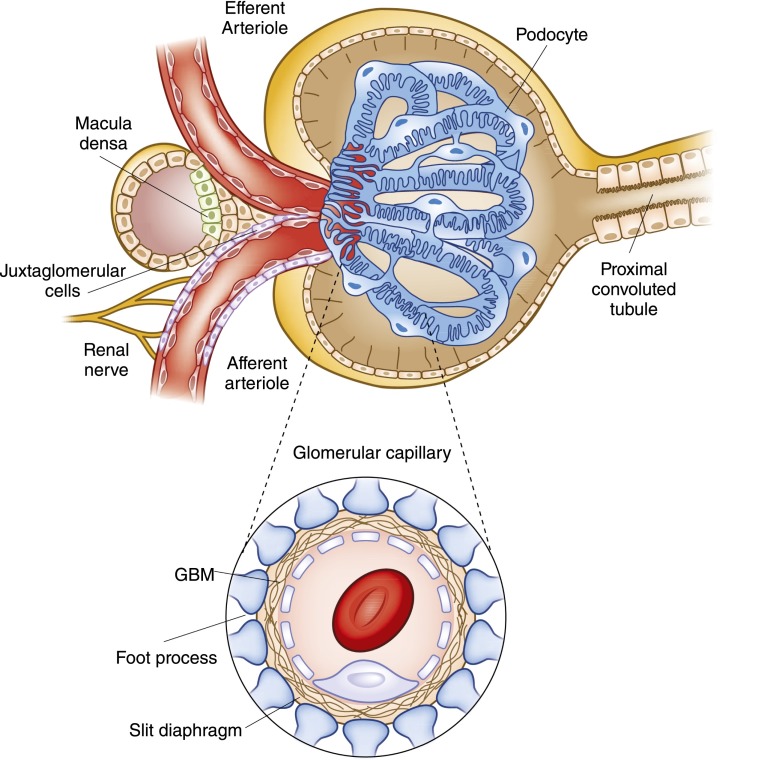
The glomerulus, the filtering unit of the kidney, is a unique bundle of capillaries lined by delicate fenestrated endothelia, a complex mesh of proteins that serve as the glomerular basement membrane and specialized visceral epithelial cells that form the slit diaphragms between interdigitating foot processes. Taken together, this arrangement allows continuous filtration of the plasma volume. The dynamic physical forces that determine the single nephron glomerular filtration are considered. In addition, new insights into the cellular and molecular components of the glomerular tuft and their contribution to glomerular disorders are explored.
Keywords: glomerular filtration; glomerulus; renal physiology.
Copyright © 2014 by the American Society of Nephrology.
Figures
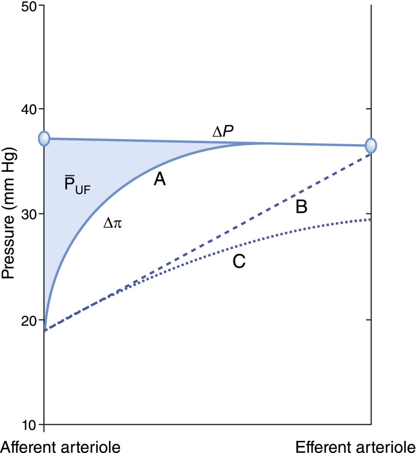
 is equal to the shaded region between the
  and the
  curves. Curve B is a hypothetical linear profile of the oncotic pressure that would occur with a minimum value of the Kf. By contrast, in curve C, filtration equilibrium is never reached. Modified from Taal MW, Chertow GM, Marsden PA, Skorecki K, Yu ASL, Brenner BM: Brenner and Rector's The Kidney, 9th Ed., Philadelphia, Elsevier Saunders, 2012, with permission.
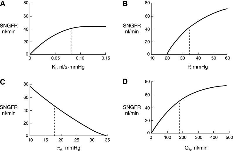
 = 35 mmHg,
  = 18 mmHg, and QA = 135 nl/min. (A) Alterations in Kf; (B) alterations in
  ; (C) alterations in ; (D) alterations in QA. Modified from Brenner BM, Dworkin LD, Ichikawa I: Glomerular filtration. In: Brenner and Rector’s The Kidney, 3rd Ed., edited by Brenner BM, Rector FC Jr, Philadelphia, Elsevier, 1986, with permission.
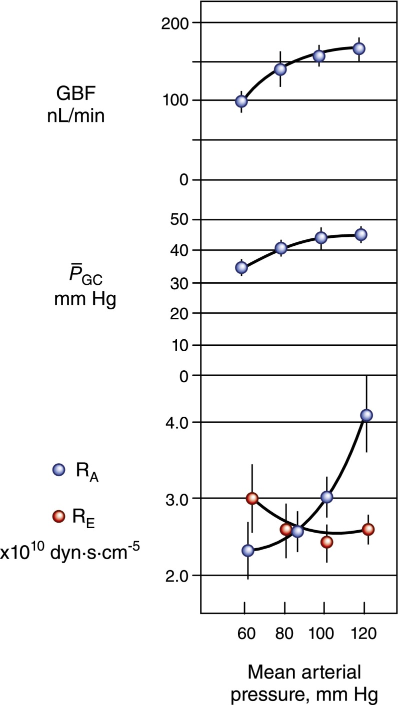
 remained relatively constant over the range of pressure from 80 to 120 mmHg, in response to a marked decrease in afferent arteriolar resistance (RA). With further reduction in perfusion pressure to 60 mmHg, GBF declined proportionally more than
  primarily because of an increase in RE. Reprinted from Taal MW, Chertow GM, Marsden PA, Skorecki K, Yu ASL, Brenner BM: Brenner and Rector’s The Kidney, 9th Ed., Philadelphia, Elsevier Saunders, 2012, with permission.
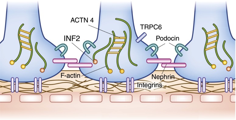
References
-
- Hinchliffe SA, Sargent PH, Howard CV, Chan YF, van Velzen D: Human intrauterine renal growth expressed in absolute number of glomeruli assessed by the disector method and Cavalieri principle. Lab Invest 64: 777–784, 1991 - PubMed
-
- Horster M, Thurau K: Micropuncture studies on the filtration rate of single superficial and juxtamedullary glomeruli in the rat kidney. Pflugers Arch Gesamte Physiol Menschen Tiere 301: 162–181, 1968 - PubMed
-
- Gong R, Dworkin LD, Brenner BM, Maddox DA: The renal circulations and glomerular ultrafiltration. In: Brenner & Rector’s The Kidney, edited by Brenner BM, Philadelphia, Elsevier Saunders, 2008, pp 91–129
-
- Stein JH, Boonjarern S, Wilson CB, Ferris TF: Alterations in intrarenal blood flow distribution. Methods of measurement and relationship to sodium balance. Circ Res 32[Suppl 1]: 61–72, 1973 - PubMed
Publication types
MeSH terms
LinkOut - more resources
Full Text Sources
Other Literature Sources
Medical
