Review
. 2014 Aug;28(8):915-30.
doi: 10.1038/eye.2014.110. Epub 2014 May 30.

The impact of new methods of investigation and treatment on the understanding of the pathology of scleral inflammation

Affiliations
Review

The impact of new methods of investigation and treatment on the understanding of the pathology of scleral inflammation

P Watson et al. Eye (Lond). 2014 Aug.

Abstract

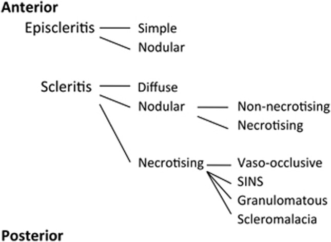
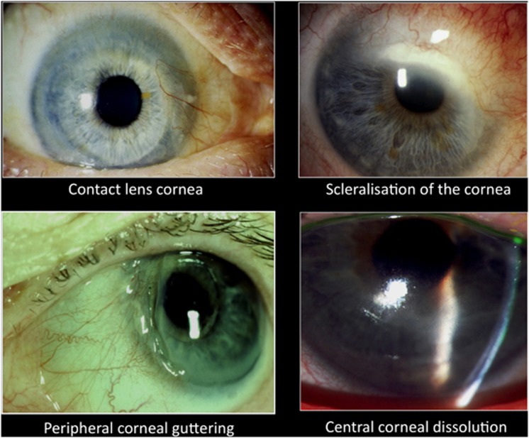
Recent advances in the understanding of the initiation and perpetuation of the immune response strongly suggest that all forms of noninfective immunologically induced scleral inflammation have a common origin. Analysis of the progress of patients with scleritis corroborates the current clinical classification that, together with studies of the immunohistology fluoresceine/ICG angiography, 3D proteoglycan, and keratan sulphate electron microscopy of scleritis, strongly suggests that from the initiation of the inflammatory process, necrotizing scleritis and diffuse and nodular scleritis not only pursue a different course but also have a different pathogenesis; nonnecrotizing scleritis being the consequence of an auto immune response, whereas necrotizing scleritis being the complication of an already present (if not always manifest), systemic immune-mediated systemic disease and its associated vasculitis. The increasing imaging capacity of anterior segment ocular coherence tomography (OCT) and en face OCT enables the changes occurring in the sclera during the course of the disease to be observed for the first time. These observations suggest that the inflammatory changes involve the potential suprachoroidal space between choroid and sclera, an observation supported by the presence of subscleral granulomas on histopathology. New imaging techniques have also been able to explain the changes seen in the cornea as a complication of scleritis. These findings have implications for investigation and the treatment of these conditions.


Figures

Figure 1
Figure 1
The current classification of noninfective scleral inflammation modified from that proposed in 1968 (see ref. pp 154–160). Apart from the additional differentiation between the nonnecrotizing and the necrotizing form of nodular scleritis, this classification remains correct. Posterior scleritis is nonnecrotizing even when the choroid is also affected. SINS is scleritis following trauma often surgically induced.
Figure 2
Figure 2
Normal anterior sclera, cornea, and chamber angle as seen with the new algorithm for the anterior segemnt OCT. SS, scleral spur; SC, Schlemm's canal; SL, Schwalbe's line.
Figure 3
Figure 3
Diffuse anterior scleritis. Anterior segment OCT. There is marked oedmea of the sclera with separation of the collagen fibres and infitration with inflammatory cells in the deeper layers of the sclera. BV, blood vessels.
Figure 4
Figure 4
Nonnecrotizing nodular scleritis. Anterior segment OCT. The nodule consists of extracellular fluid. The collagen fibres are separated but remain distinct. There is no necrosis of tissue.
Figure 5
Figure 5
Histology of a nonnecrotizing scleral nodule. The inflammation affects the whole thickness of the sclera.
Figure 6
Figure 6
Necrotizing nodular scleritis. Anterior segment OCT. There is hyperreflectivity of the episcleral and deep tissues. The hyporeflectivity of the scleral nodule shows that the deep layers of the nodule are liquefied and interpersed with blood vessels.
Figure 7
Figure 7
Necrotizing scleritis. Apart from the obvious loss of tissue the collagen fibres are aggregated together, indicating a loss of the proteoglycan coat. There is not only erosion of the superficial tissues but also under surface of the sclera.
Figure 8
Figure 8
En face OCT in necrotizing scleritis. The depth, structure, and extent of the lesion can be determined. En face image integrated over the full-scan range of the sclera. Horizontal B-scan image through the centre of the scleral lesion in a patient with necrotizing scleritis (i) with overlay contour lines indicating the positions of partial intensity en face images. En face image of the 30 μm thick slab immediately underneath the conjunctival epithelium (a–h). A hyporeflective image indicates destruction of the posterior surface of the sclera (f–h).
Figure 9
Figure 9
Scleral collagen and its associated proteoglycans. Scleral collagen fibrils exhibit a–e cross-banding in longitudinal sections (top panel). Cuprolinic blue staining visualizes proteglycans as filaments is associated with the d bands. These filaments lie along the fibril, encircle it, and connect the collagen fibrils to each other (lower panel).
Figure 10
Figure 10
The distribution of immume proteins in cornea and sclera.
Figure 11
Figure 11
(Left above) Zonal necrotizing granulomatous inflammation in rheumatoid arthritis showing an acellular necrotic centre with an infiltration of inflammatory cells and some giant cells. (Left below) Staining for CD20 B cells in the same patient. (Right above) Scleritis in a patient with no evidence of systemic disease. There is diffuse infiltration of the sclera with inflammatory cells. (Right below) Staining for CD8 macrophages in the top right patient. The predominant cell is the macrophage (ref. p 177, Figure 7.39).
Figure 12
Figure 12
Granulomatous scleritis in granulomatosis with polyangiitis (Wegener's granulomatosis). (a) The characteristic presentation where the inflammation includes the limbus and adjacent cornea (in rheumatoid arthritis the limbal area is spared). (b) Red Free and ICG after 2 weeks of intensive treatment. The eye appears completely quiet on the red free but ICG reveals an intense staining at an area of vasculitis. Treatment should not be discontinued until these areas have healed.
Figure 13
Figure 13
Necrotizing scleritis associated with granulomatosis with polyangiitis (Wegener's granulomatosis). A 57-year-old woman who presented with a red eye that was painful for 3 months. (a) The corneal destructive changes involve the whole limbus typical of that seen in a systemic vasculitis even though the ANCA was negative at this time. (b) After 17 months. Advice that she should be treated systemically was not accepted even though she had become ANCA positive by April 2001.
Figure 14
Figure 14
Normal collagen/proteoglycan structure on human sclera. In necrotising scleritis the proteoglycan separates from the collagen leading to unravelling of the collagen and its exposure to cytokine activity. (a) Proteoglycan fibrils connecting collagen to collagen. (b) Encircling proteoglycan fibril. (c) Proteoglycan fibril along the collagen (ref. pp 158-159, Figures 7.10, 7.11, 7.12, 7.13).
Figure 15
Figure 15
The corneal changes associated with scleral inflammation.
Figure 16
Figure 16
Contact lens cornea. Fluoresceine/ICG angiograms showing closure of the episleral capillary network associated with long-standing scleral inflammation. Fluorescein shows closure (arrow) with secondary overgrowth of the conjunctival vessels over the limbus to the edge of cornea of normal thickness. ICG images confirm the gross reduction of episcleral capillaries and leakage from the remaining ones (between arrows).
Figure 17
Figure 17
Cross-sectional and longitudinal TEM micrographs of collagen fibrils across the bovine cornea and sclera. The collagen fibril diameter is uniform from the cornea centre until the limbus where it varies to increase significantly into the sclera. This is so in the outer mid and deep stroma. Scale bar=100 nm (cross-sectional) and 200 nm (longitudinal). The mean fibril diameter across the bovine cornea and sclera is shown in the graph. 0–3 nm=centre; 3–6 nm=inner perphery; 6–9=mid periphery, 9–12=outer periphery; and 12–15=sclera (Ho et al, submitted).
Figure 18
Figure 18
The ‘stitched' composite fundus photgraph taken with an attachment to a smart phone of an exudative retinal detachment and retinal haemorrhages taken by a health-care worker in the PEEK project in Kenya ready to be transmitted to a diagnostic centre.
Figure 19
Figure 19
Masai health-care worker in PEEK project. He has two watches and his diagnostic smart phone in his belt.

References

    1. Wiiliamson J. Incidence of eye disease in cases of connective tissue disease. Trans Ophthal Soc UK. 1974;94:742–752. - PubMed
    1. Watson PG, Hazleman BL, McKluskey P, Pavesio CE.The Sclera and Systemic Disorders3rd edn.JP Medical: London; 2012
    1. Sainz de la Maza M, Tauber J, Foster CS.The Sclera2nd ednSpringer Verlag: New York; 2012
    1. Wieringa WG, Wieringa JE, ten Dam-van Loon NH, Los LI. Visual outcome, treatment results, and prognostic factors in patients with scleritis. Ophthalmology. 2013;120 (2:379–386. - PubMed
    1. Watson PG, Young RD. Scleral structure, organisation and disease. Exp Eye Res. 2004;78 (3:609–623. - PubMed