. 2014;13(14):2248-61.
doi: 10.4161/cc.29270. Epub 2014 May 29.

Spatial regulation of Aurora A activity during mitotic spindle assembly requires RHAMM to correctly localize TPX2

Affiliations

Spatial regulation of Aurora A activity during mitotic spindle assembly requires RHAMM to correctly localize TPX2

Helen Chen et al. Cell Cycle. 2014.

Abstract

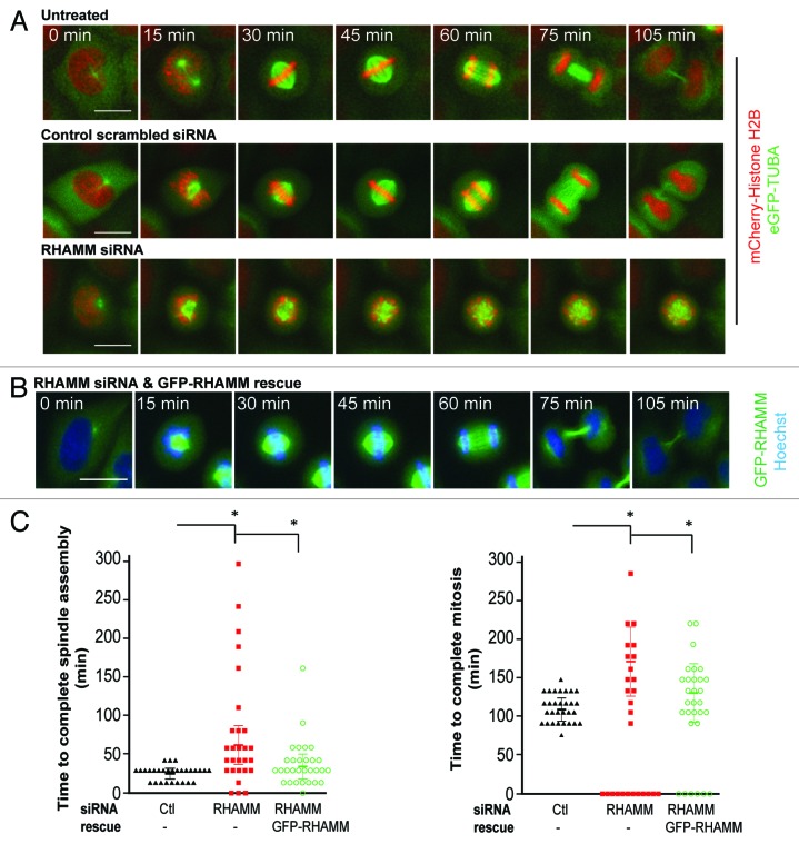
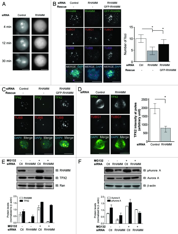
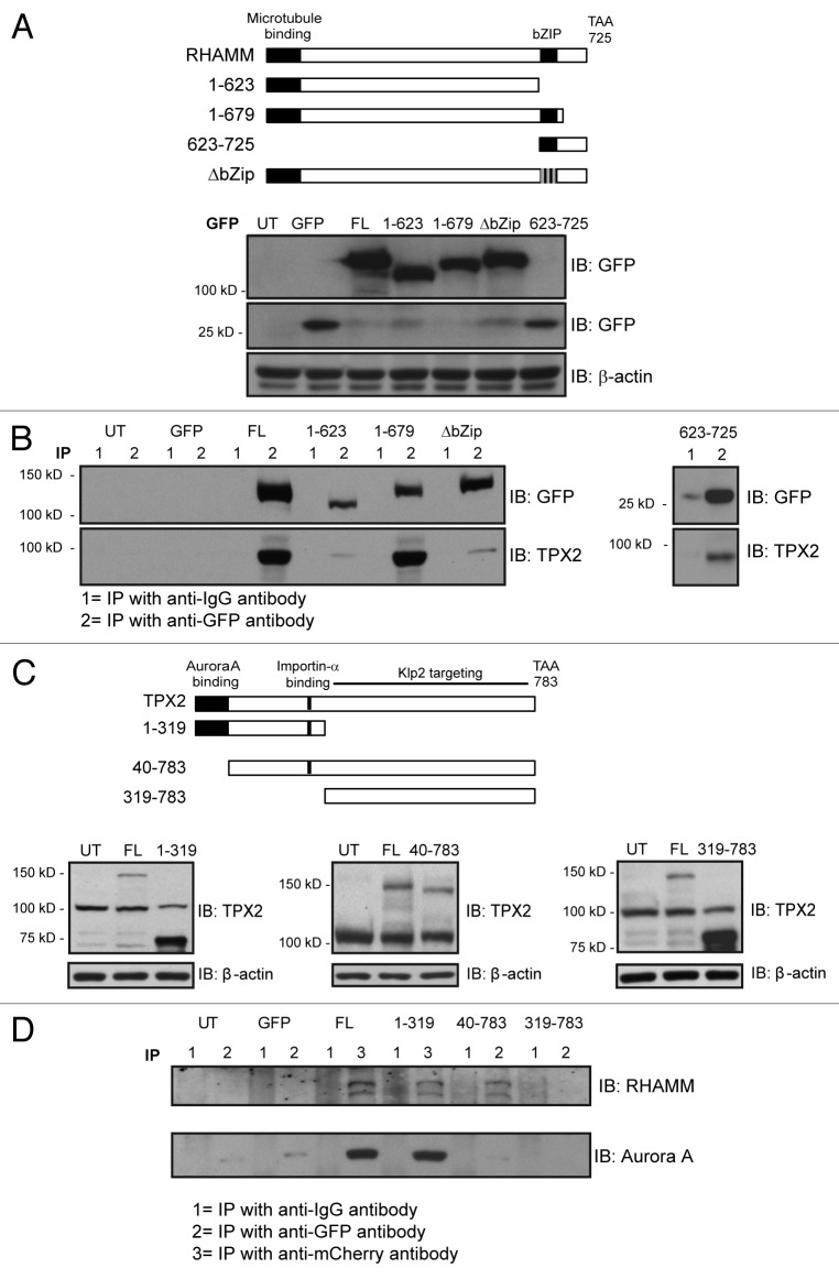
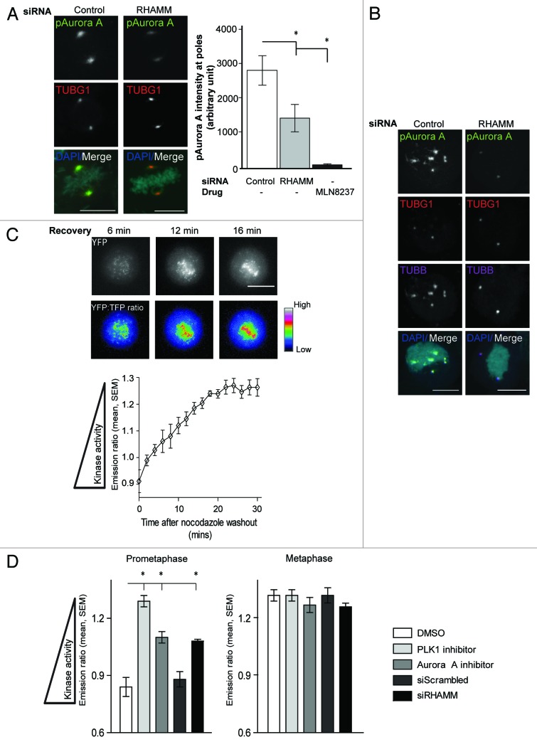
Construction of a mitotic spindle requires biochemical pathways to assemble spindle microtubules and structural proteins to organize these microtubules into a bipolar array. Through a complex with dynein, the receptor for hyaluronan-mediated motility (RHAMM) cross-links mitotic microtubules to provide structural support, maintain spindle integrity, and correctly orient the mitotic spindle. Here, we locate RHAMM to sites of microtubule assembly at centrosomes and non-centrosome sites near kinetochores and demonstrate that RHAMM is required for the activation of Aurora kinase A. Silencing of RHAMM delays the kinetics of spindle assembly, mislocalizes targeting protein for XKlp2 (TPX2), and attenuates the localized activation of Aurora kinase A with a consequent reduction in mitotic spindle length. The RHAMM-TPX2 complex requires a C-terminal basic leucine zipper in RHAMM and a domain that includes the nuclear localization signal in TPX2. Together, our findings identify RHAMM as a critical regulator for Aurora kinase A signaling and suggest that RHAMM ensures bipolar spindle assembly and mitotic progression through the integration of biochemical and structural pathways.

Keywords: Aurora kinase A; RHAMM; TPX2; mitosis; spindle assembly.


Figures

None
Figure 1. RHAMM participates in spindle microtubule assembly at both centrosomal and non-centrosomal sites during early mitosis and is required for proper spindle architecture and mitosis kinetics. (A) In prophase HeLa cells, RHAMM was located at the centrosomes (identified by gamma-tubulin, TUBG1) as well as non-centrosomal sites near kinetochores (identified by BubR1) within nuclear volumes (identified by DAPI) as determined for endogenous RHAMM by immunofluorescence and by the expression of exogenous GFP-RHAMM. Scale bars = 10 μm. (B) In prophase RPMI 8226 cells, exogenous GFP-RHAMM colocalized with TUBG1 at the centrosomes and BubR1 at the kinetochores within nuclear volumes. Scale bars = 10 μm. (C) In mitotic HeLa cells, RHAMM localization at spindle microtubule assembly sites was tracked at indicated times following depolymerization by nocodazole. Microtubule assembly (identified by β-tubulin, TUBB) initiated at 2 min around the centrosomes (TUBG1) and included non-centrosomal sites near the kinetochores (BubR1) by 6 min. RHAMM was located at both centrosomes and non-centrosomal spindle microtubule assembly sites. Scale bars = 10 μm. (D) HeLa cells were treated with scrambled control (Ctl) siRNA or siRNA duplexes targeting the 3′ UTR (#1, #2) or the 5′ UTR (#3) of RHAMM mRNA as well as pooled siRNA (1–3) to deplete endogenous RHAMM. RHAMM expression was measured by western blot analysis 48–72 h following transfection. Equal loading was confirmed with GAPDH. (E) Exogenous GFP-RHAMM was transfected into cells 48 h after siRNA transfection. After a further 48 h, the expression of RHAMM, and GFP-RHAMM (shifted by 27 kD), were measured by western blot analysis. GFP-RHAMM was expressed in RHAMM-silenced cells at a level similar to that of endogenous RHAMM. Equal loading was confirmed with GAPDH. (F) Aberrant spindle figures (multipolar spindle, disorganized spindle and unattached chromosomes) were significantly more frequent in RHAMM-silenced cells. Rescue with GFP-RHAMM was sufficient to reduce these aberrant phenotypes. (mean ± s.d., n = 3, *P < 0.05). (G) Multinucleated cells were significantly more frequent in RHAMM-silenced cells. Rescue with GFP-RHAMM was sufficient to reduce these aberrant phenotypes. (mean ± s.d., n = 3, *P < 0.05). (H) Mitotic indices were determined by DAPI staining in immunofluorescence. The mitotic index was significantly higher in RHAMM-silenced cells. Rescue with GFP-RHAMM was sufficient to reduce the mitotic index. (mean ± s.d., n = 3, *P < 0.05)
None
Figure 2. Proper mitotic kinetics and spindle microtubule assembly are dependent on RHAMM expression. (A) Living HeLa cells expressing eGFP-tubulin and mCherry Histone-H2B were treated with either control siRNA or siRNA duplexes targeting RHAMM and followed through mitosis using time-lapse microscopy. Scale bars = 10 μm. (B) HeLa cells were treated with siRNA duplexes targeting RHAMM and rescued with exogenous GFP-RHAMM. Cells were labeled with Hoechst to visualize DNA and followed through mitosis using time-lapse microscopy. Scale bars = 10 μm. (C) The kinetics for mitosis in RHAMM-silenced cells was delayed. Expression of exogenous GFP-RHAMM significantly reduced the time to transit through mitosis. Cells that did not complete mitosis during imaging are plotted at 0 min and were not included in the quantitation of mitosis duration. (mean ± s.d., n = 6, *P < 0.05).
None
Figure 3. RHAMM is required for spindle microtubule assembly and TPX2 localization and stability. (A) In living HeLa cells expressing GFP-tubulin, microtubule assembly and the formation of a bipolar spindle following nocodazole treatment was disrupted in cells treated with siRNA targeting RHAMM. Scale bars = 10 μm. (B) In mitotic HeLa cells recovering from nocodazole treatment, microtubule assembly at non-centrosomal sites was attenuated in RHAMM-silenced cells, and the expression of GFP-RHAMM was sufficient to re-establish microtubule assembly at these sites. Scale bars = 10 μm. (mean ± s.d., n = 4, *P < 0.05). (C) TPX2 localization to non-centrosomal microtubule assembly sites was abolished in RHAMM-silenced mitotic cells during recovery from nocodazole, and the expression of GFP-RHAMM was sufficient to re-establish microtubule assembly at these sites. Scale bars = 10 μm. (D) TPX2 specific immuno-staining at spindle poles (regions of interest identified by TUBG1) was significantly reduced in cells treated with siRNA targeting RHAMM. Scale bars = 10 μm. (mean ± s.d., n = 3, *P < 0.05). (E) In synchronized early mitotic cells, the levels of RHAMM and TPX2 were reduced in cells pretreated with siRNA targeting RHAMM compared with those treated with scrambled control siRNA. Proteasome inhibition (MG132, 15 µM) was sufficient to recover the level of TPX2 in RHAMM-silenced cells. Ran served as a related protein that was unaffected in RHAMM-silenced cells. Protein expression levels in immunoblotting experiments are quantified by normalizing to β-actin levels. (F) In synchronized early mitotic cell, the levels of Aurora A and pAurora A were reduced in cells pretreated with siRNA targeting RHAMM compared with those treated with scrambled control siRNA. Proteasome inhibition (MG132, 15 µM) was sufficient to recover the level of Aurora A, but not pAurora A, in RHAMM-silenced cells. β-actin levels confirmed equal loading. Protein expression levels in immunoblotting experiments are quantified by normalizing to β-actin levels.
None
Figure 4. Characterization of the functional domains involved in RHAMM-TPX2 interaction during early mitosis. (A) Schematic diagram of defined domains in RHAMM. The 3 gray lines in the ΔbZIP construct represent leucines mutated to arginines. Western blot analysis confirmed the expression of GFP-RHAMM constructs. β-actin levels confirmed equal loading. (B) Immunoprecipitation of GFP-RHAMM constructs identified the bZIP motif as a necessary domain in RHAMM for the co-precipitation of TPX2. Cell lysates were immunoprecipitated with either an IgG control antibody (lanes marked 1) or antibodies against eGFP (lanes marked 2). (C) Schematic diagram of defined domains in TPX2. Western blot analysis confirmed the expression of mCherry-TPX2 and TPX2-GFP truncation variants. β-actin levels confirmed equal loading. (D) Immunoprecipitation of mCherry–TPX2 or TPX2–GFP truncation variants identified amino acids 40–319 as necessary for the co-precipitation of RHAMM. Cell lysates were immunoprecipitated with either an IgG control antibody (lane marked 1) or antibodies against eGFP (lane marked 2) or mCherry (lane marked 3).
None
Figure 5. Proper mitotic spindle length requires RHAMM targeting TPX2 to the spindle poles. (A) Cells treated with siRNA targeting RHAMM were transfected with the indicated GFP-RHAMM constructs, and asynchronous metaphase cells were analyzed by immunofluorescence. Amino acids 623–679 of RHAMM are the minimal required domain for protein localization to spindle poles (identified by TUBG1), while TPX2 localization to the spindle poles require additional residues. Scale bars = 10 μm. (B) Spindle length was measured based on TUBG1 staining in cells treated with either control siRNA or RHAMM targeted siRNA and followed by rescue with indicated GFP-RHAMM constructs. Measurements are made in a 3D projection along a plane perpendicular to the chromosomes. The bZIP motif (623–679) was necessary, though not sufficient, to establish proper mitotic spindle length. (mean ± SEM, n = 3, 30 cells per treatment, *P < 0.05). (C) RHAMM remained localized at the spindle poles in cells treated with shRNA targeting TPX2. Scale bars = 10 μm. (D) Cells treated with shRNA targeting TPX2 were transfected with the indicated mCherry-TPX2 or TPX2-GFP constructs and asynchronous metaphase cells were analyzed by immunofluorescence. Scale bars = 10 μm. (E) Spindle length was measured based on TUBG1 staining in cells treated with either control shNHP or TPX2 targeted shRNA, and followed by rescue with indicated mCherry–TPX2 or TPX2–GFP constructs. Measurements are made in a 3D projection along a plane perpendicular to the chromosomes. (mean ± SEM, n = 1, >15 cells per treatment, *P < 0.05)
None
Figure 6. RHAMM is required for the temporally- and spatially- regulated activity of Aurora A. (A) pAurora A specific immunoflourescence at the spindle poles (regions of interest identified by TUBG1) was significantly reduced in cells treated with MLN8237 (Aurora A specific inhibitor) or siRNA targeting RHAMM, relative to control siRNA-treated cells. Scale bars = 10 μm. (mean ± s.d., n = 3, *P < 0.05). (B) pAurora A localization to non-centrosome microtubule assembly sites was abolished in RHAMM-silenced mitotic cells during recovery from nocodazole treatment. Scale bars = 10 μm. (C) HeLa cells expressing a Hec-1 targeted PLK1 FRET sensor were imaged during recovery from nocodazole treatment. Phosphorylation of the kinetochore-targeted sensor was highest in early prometaphase (0–18 min) and diminished to background levels by metaphase (18–20 min) as indicated by the YFP:TFP emission ratio. Scale bars = 10 μm. (mean ± SEM., n = 3, 6 cells per treatment). (D) Relative to measurements in cells incubated with DMSO vehicle control, FRET ratios for the Hec-1 targeted PLK1 substrate were significantly higher in cells incubated with either a PLK1-specific inhibitor (BI2536, 20 nM for 2 h) or an Aurora A-specific inhibitor (MLN8237, 1 µM for 2 h) during prometaphase (2 min after nocodazole washout). Pretreatment of cells with siRNA targeting RHAMM, but not with scrambled siRNA controls, also significantly augmented the FRET emission ratio in prometaphase cells. No significant change in the FRET ratio was observed in metaphase cells (20 min after nocodazole washout) (mean ± SEM., n = 3, 6 cells per treatment, *P < 0.05).

Similar articles

Cited by

References

    1. Walczak CE, Cai S, Khodjakov A. Mechanisms of chromosome behaviour during mitosis. Nat Rev Mol Cell Biol. 2010;11:91–102. - PMC - PubMed
    1. Barros TP, Kinoshita K, Hyman AA, Raff JW. Aurora A activates D-TACC-Msps complexes exclusively at centrosomes to stabilize centrosomal microtubules. J Cell Biol. 2005;170:1039–46. doi: 10.1083/jcb.200504097. - DOI - PMC - PubMed
    1. Giet R, McLean D, Descamps S, Lee MJ, Raff JW, Prigent C, Glover DM. Drosophila Aurora A kinase is required to localize D-TACC to centrosomes and to regulate astral microtubules. J Cell Biol. 2002;156:437–51. doi: 10.1083/jcb.200108135. - DOI - PMC - PubMed
    1. Terada Y, Uetake Y, Kuriyama R. Interaction of Aurora-A and centrosomin at the microtubule-nucleating site in Drosophila and mammalian cells. J Cell Biol. 2003;162:757–63. doi: 10.1083/jcb.200305048. - DOI - PMC - PubMed
    1. Mori D, Yano Y, Toyo-oka K, Yoshida N, Yamada M, Muramatsu M, Zhang D, Saya H, Toyoshima YY, Kinoshita K, et al. NDEL1 phosphorylation by Aurora-A kinase is essential for centrosomal maturation, separation, and TACC3 recruitment. Mol Cell Biol. 2007;27:352–67. doi: 10.1128/MCB.00878-06. - DOI - PMC - PubMed

Publication types

MeSH terms

LinkOut - more resources