Enriched endogenous omega-3 fatty acids in mice protect against global ischemia injury
- PMID: 24875538
- PMCID: PMC4076077
- DOI: 10.1194/jlr.M046466
Enriched endogenous omega-3 fatty acids in mice protect against global ischemia injury
Abstract
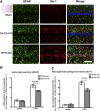
Transient global cerebral ischemia, one of the consequences of cardiac arrest and cardiovascular surgery, usually leads to delayed death of hippocampal cornu Ammonis1 (CA1) neurons and cognitive deficits. Currently, there are no effective preventions or treatments for this condition. Omega-3 (ω-3) PUFAs have been shown to have therapeutic potential in a variety of neurological disorders. Here, we report that the transgenic mice that express the fat-1 gene encoding for ω-3 fatty acid desaturase, which leads to an increase in endogenous ω-3 PUFAs and a concomitant decrease in ω-6 PUFAs, were protected from global cerebral ischemia injury. The results of the study show that the hippocampal CA1 neuronal loss and cognitive deficits induced by global ischemia insult were significantly less severe in fat-1 mice than in WT mice controls. The protection against global cerebral ischemia injury was closely correlated with increased production of resolvin D1, suppressed nuclear factor-kappa B activation, and reduced generation of pro-inflammatory mediators in the hippocampus of fat-1 mice compared with WT mice controls. Our study demonstrates that fat-1 mice with high endogenous ω-3 PUFAs exhibit protective effects on hippocampal CA1 neurons and cognitive functions in a global ischemia injury model.
Copyright © 2014 by the American Society for Biochemistry and Molecular Biology, Inc.
Figures


Similar articles
-
Enriched Endogenous Omega-3 Polyunsaturated Fatty Acids Protect Cortical Neurons from Experimental Ischemic Injury.Mol Neurobiol. 2016 Nov;53(9):6482-6488. doi: 10.1007/s12035-015-9554-y. Epub 2015 Nov 26. Mol Neurobiol. 2016. PMID: 26611833
-
Omega-3 fatty acids protect the brain against ischemic injury by activating Nrf2 and upregulating heme oxygenase 1.J Neurosci. 2014 Jan 29;34(5):1903-15. doi: 10.1523/JNEUROSCI.4043-13.2014. J Neurosci. 2014. PMID: 24478369 Free PMC article.
-
Enriched endogenous n-3 polyunsaturated fatty acids alleviate cognitive and behavioral deficits in a mice model of Alzheimer's disease.Neuroscience. 2016 Oct 1;333:345-55. doi: 10.1016/j.neuroscience.2016.07.038. Epub 2016 Jul 27. Neuroscience. 2016. PMID: 27474225
-
Improved outcome after peripheral nerve injury in mice with increased levels of endogenous ω-3 polyunsaturated fatty acids.J Neurosci. 2012 Jan 11;32(2):563-71. doi: 10.1523/JNEUROSCI.3371-11.2012. J Neurosci. 2012. PMID: 22238091 Free PMC article.
-
Resolvin D2 protects against cerebral ischemia/reperfusion injury in rats.Mol Brain. 2018 Feb 13;11(1):9. doi: 10.1186/s13041-018-0351-1. Mol Brain. 2018. PMID: 29439730 Free PMC article.
Cited by
-
The Erythrocyte Fatty Acid Profile in Multiple Sclerosis Is Linked to the Disease Course, Lipid Peroxidation, and Dietary Influence.Nutrients. 2025 Mar 11;17(6):974. doi: 10.3390/nu17060974. Nutrients. 2025. PMID: 40290001 Free PMC article.
-
n-3 Polyunsaturated Fatty Acids and Their Derivates Reduce Neuroinflammation during Aging.Nutrients. 2020 Feb 28;12(3):647. doi: 10.3390/nu12030647. Nutrients. 2020. PMID: 32121189 Free PMC article. Review.
-
Enriched Brain Omega-3 Polyunsaturated Fatty Acids Confer Neuroprotection against Microinfarction.EBioMedicine. 2018 Jun;32:50-61. doi: 10.1016/j.ebiom.2018.05.028. Epub 2018 Jun 5. EBioMedicine. 2018. PMID: 29880270 Free PMC article.
-
Protection against Oxygen-Glucose Deprivation/Reperfusion Injury in Cortical Neurons by Combining Omega-3 Polyunsaturated Acid with Lyciumbarbarum Polysaccharide.Nutrients. 2016 Jan 13;8(1):41. doi: 10.3390/nu8010041. Nutrients. 2016. PMID: 26771636 Free PMC article.
-
RvD1binding with FPR2 attenuates inflammation via Rac1/NOX2 pathway after neonatal hypoxic-ischemic injury in rats.Exp Neurol. 2019 Oct;320:112982. doi: 10.1016/j.expneurol.2019.112982. Epub 2019 Jun 24. Exp Neurol. 2019. PMID: 31247196 Free PMC article.
References
-
- Harukuni I., Bhardwaj A. 2006. Mechanisms of brain injury after global cerebral ischemia. Neurol. Clin. 24: 1–21 - PubMed
-
- Hossmann K. A. 2008. Cerebral ischemia: models, methods and outcomes. Neuropharmacology. 55: 257–270 - PubMed
-
- Liou A. K., Clark R. S., Henshall D. C., Yin X. M., Chen J. 2003. To die or not to die for neurons in ischemia, traumatic brain injury and epilepsy: a review on the stress-activated signaling pathways and apoptotic pathways. Prog. Neurobiol. 69: 103–142 - PubMed
MeSH terms
Substances
LinkOut - more resources
Full Text Sources
Other Literature Sources
Molecular Biology Databases
Miscellaneous

