

Asthmatic airway epithelial cells differentially regulate fibroblast expression of extracellular matrix components
- PMID: 24875618
- PMCID: PMC4149938
- DOI: 10.1016/j.jaci.2014.04.007
Asthmatic airway epithelial cells differentially regulate fibroblast expression of extracellular matrix components
Abstract
Background: Airway remodeling might explain lung function decline among asthmatic children. Extracellular matrix (ECM) deposition by human lung fibroblasts (HLFs) is implicated in airway remodeling. Airway epithelial cell (AEC) signaling might regulate HLF ECM expression.
Objectives: We sought to determine whether AECs from asthmatic children differentially regulate HLF expression of ECM constituents.
Methods: Primary AECs were obtained from well-characterized atopic asthmatic (n = 10) and healthy (n = 10) children intubated during anesthesia for an elective surgical procedure. AECs were differentiated at an air-liquid interface for 3 weeks and then cocultured with HLFs from a healthy child for 96 hours. Collagen I (COL1A1), collagen III (COL3A1), hyaluronan synthase (HAS) 2, and fibronectin expression by HLFs and prostaglandin E2 synthase (PGE2S) expression by AECs were assessed by using RT-PCR. TGF-β1 and TGF-β2 concentrations in media were measured by using ELISA.
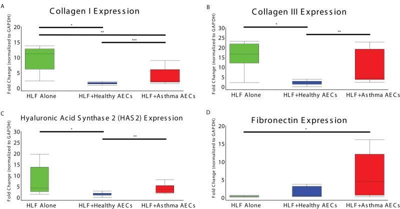
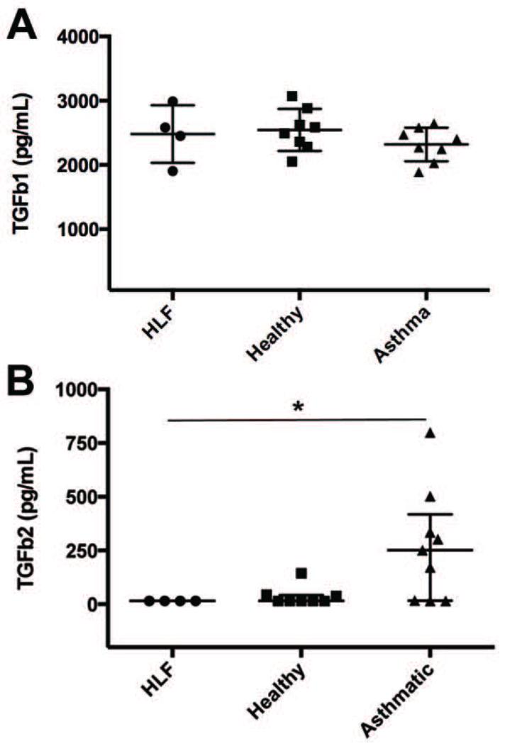
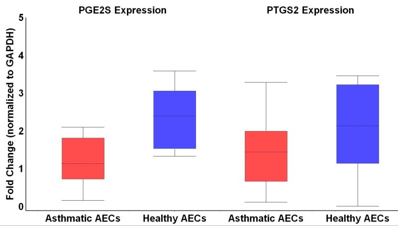
Results: COL1A1 and COL3A1 expression by HLFs cocultured with AECs from asthmatic patients was greater than that by HLFs cocultured with AECs from healthy subjects (2.2-fold, P < .02; 10.8-fold, P < .02). HAS2 expression by HLFs cocultured with AECs from asthmatic patients was 2.5-fold higher than that by HLFs cocultured with AECs from healthy subjects (P < .002). Fibronectin expression by HLFs cocultured with AECs from asthmatic patients was significantly greater than that by HLFs alone. TGF-β2 activity was increased in cocultures of HLFs with AECs from asthmatic patients (P < .05), whereas PGES2 was downregulated in AEC-HLF cocultures (2.2-fold, P < .006).
Conclusions: HLFs cocultured with AECs from asthmatic patients showed differential expression of the ECM constituents COL1A1 and COL3A1 and HAS2 compared with HLFs cocultured with AECs from healthy subjects. These findings support a role for altered ECM production in asthmatic airway remodeling, possibly regulated by unbalanced AEC signaling.
Keywords: Asthma; TGF-β2; airway remodeling; children; collagen I; collagen III; epithelial cells; extracellular matrix; fibronectin; human lung fibroblasts; hyaluronic acid.
Copyright © 2014 American Academy of Allergy, Asthma & Immunology. Published by Elsevier Inc. All rights reserved.
Figures
Similar articles
-
Fibroblast gene expression following asthmatic bronchial epithelial cell conditioning correlates with epithelial donor lung function and exacerbation history.Sci Rep. 2018 Oct 25;8(1):15768. doi: 10.1038/s41598-018-34021-6. Sci Rep. 2018. PMID: 30361541 Free PMC article.
-
Fibroblast-myofibroblast transition is differentially regulated by bronchial epithelial cells from asthmatic children.Respir Res. 2015 Feb 13;16(1):21. doi: 10.1186/s12931-015-0185-7. Respir Res. 2015. PMID: 25849331 Free PMC article.
-
Asthmatic bronchial epithelial cells promote the establishment of a Hyaluronan-enriched, leukocyte-adhesive extracellular matrix by lung fibroblasts.Respir Res. 2018 Aug 2;19(1):146. doi: 10.1186/s12931-018-0849-1. Respir Res. 2018. PMID: 30071849 Free PMC article.
-
The Role of miRNAs in Extracellular Matrix Repair and Chronic Fibrotic Lung Diseases.Cells. 2021 Jul 6;10(7):1706. doi: 10.3390/cells10071706. Cells. 2021. PMID: 34359876 Free PMC article. Review.
-
Extracellular Matrix Component Remodeling in Respiratory Diseases: What Has Been Found in Clinical and Experimental Studies?Cells. 2019 Apr 11;8(4):342. doi: 10.3390/cells8040342. Cells. 2019. PMID: 30979017 Free PMC article. Review.
Cited by
-
Drugs Used in the Treatment of Viral Infections for the Prevention of Airway Remodeling in Asthma.Mediators Inflamm. 2025 Jul 24;2025:5526526. doi: 10.1155/mi/5526526. eCollection 2025. Mediators Inflamm. 2025. PMID: 40746413 Free PMC article. Review.
-
A Heterotopic Xenograft Model of Human Airways for Investigating Fibrosis in Asthma.Am J Respir Cell Mol Biol. 2017 Mar;56(3):291-299. doi: 10.1165/rcmb.2016-0065MA. Am J Respir Cell Mol Biol. 2017. PMID: 27788019 Free PMC article.
-
Disease Models: Lung Models for Testing Drugs Against Inflammation and Infection.Handb Exp Pharmacol. 2021;265:157-186. doi: 10.1007/164_2020_366. Handb Exp Pharmacol. 2021. PMID: 33095300
-
Follistatin-Like Proteins: Structure, Functions and Biomedical Importance.Biomedicines. 2021 Aug 12;9(8):999. doi: 10.3390/biomedicines9080999. Biomedicines. 2021. PMID: 34440203 Free PMC article. Review.
-
Hyaluronan as a therapeutic target in human diseases.Adv Drug Deliv Rev. 2016 Feb 1;97:186-203. doi: 10.1016/j.addr.2015.10.017. Epub 2015 Nov 2. Adv Drug Deliv Rev. 2016. PMID: 26541745 Free PMC article. Review.
References
-
- Unit ALAEaS, Association. AL. American Lung Association, Epidemiology and Statistic Unit, Research and Program Services . Estimated prevalence and incidence of lung disease by Lung Association territory [electronic resource] American Lung Association; New York, NY: 2006.
-
- Bloom B, Cohen RA, Freeman G. Summary health statistics for U.S. children: National Health Interview Survey, 2007. Vital Health Stat. 2009;10:1–80. - PubMed
-
- Martinez FD, Wright AL, Taussig LM, Holberg CJ, Halonen M, Morgan WJ, The Group Health Medical Associates Asthma and wheezing in the first six years of life. N Engl J Med. 1995;332:133–8. - PubMed
-
- Phelan PD, Robertson CF, Olinsky A. The Melbourne Asthma Study: 1964-1999. J Allergy Clin Immunol. 2002;109:189–94. - PubMed
Publication types
MeSH terms
Substances
Grants and funding
LinkOut - more resources
Full Text Sources
Other Literature Sources
Miscellaneous
