. 2014 May 30:4:5088.
doi: 10.1038/srep05088.

Endogenous antibodies for tumor detection

Affiliations

Endogenous antibodies for tumor detection

Barrie S Rich et al. Sci Rep. .

Abstract

The study of cancer immunology has provided diagnostic and therapeutic instruments through serum autoantibody biomarkers and exogenous monoclonal antibodies. While some endogenous antibodies are found within or surrounding transformed tissue, the extent to which this exists has not been entirely characterized. We find that in transgenic and xenograft mouse models of cancer, endogenous gamma immunoglobulin (IgG) is present at higher concentration in malignantly transformed organs compared to non-transformed organs in the same mouse or organs of cognate wild-type mice. The enrichment of endogenous antibodies within the malignant tissue provides a potential means of identifying and tracking malignant cells in vivo as they mutate and diversify. Exploiting these antibodies for diagnostic and therapeutic purposes is possible through the use of agents that bind endogenous antibodies.


Figures
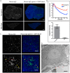
Figure 1
Figure 1. In the alb-myc transgenic model of hepatocellular carcinoma, endogenous antibodies are identified at significantly higher levels than in matched wild-type controls.
(A). Liver tissue stained with anti-mouse IgG labeled with AlexaFluor 488 viewed at low magnification demonstrates more antibody in tumor tissue than in normal wild type (WT) liver (scale bars = 1 mm). (B). Integrated intensity along the top 60% of intensity values demonstrates 28-fold greater fluorescence in tumor tissue than WT (p = 0.0002). (C). Confocal slices through carcinoma tissue reveal two distinct staining patterns: staining of sinusoidal endothelial cells (yellow arrows); and staining of hepatocytes (red arrows) with the staining in the cytosol, but excluded from the nucleus, consistent with endocytosis of the antibody (scale bars = 50 microns). (D). Binding to sinusoidal endothelial cells, arrows, was confirmed with immuno-electron microscopy (scale bar = 2 um).
Figure 2
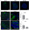
Figure 2. MMTV-neu is a transgenic model of murine mammary cancer.
(A). Staining for endogenous antibody within the neoplastic tissue demonstrated increased overall fluorescence compared to wild type mammary tissue (scale bar = 1 mm). (B). On confocal microscopy, the fluorescent signal corresponded to intraluminal glandular regions as well as the stroma (scale bar = 50 um). (C). Fluorescence labeling of mouse IgG in neoplastic mammary tissue is statistically higher than in wild type mammary tissue (p = 0.001).
Figure 3
Figure 3
(A). Livers from Alb/c-myc mice were graded according to degree of dysplasia. Bar graph represents integrated fluorescent intensity for WT tissue and graded transgenic tissue. All grades were significantly brighter than WT tissue. (grade 4: p = 0.04, grade 3: p = 0.0003, grade 2: p = 0.0007, grade 1: p = 0.01, grade 0: p = 0.0002). (B). Organs from transgenic MMTV-neu mice were stained with anti-mouse antibody to determine the specificity of the endogenous antibodies to the tumor-bearing organ. Results are shown as ratios of the fluorescence from transgenic organ to wild type organ for colon, kidney, lung, brain, heart, breast, liver, spleen, and stomach.
Figure 4
Figure 4
(A). In two transgenic models of prostate cancer, more endogenous antibody was identified in tumor bearing tissue than wild type when viewed at low magnification. (B). High magnification confocal images demonstrate antibody localization. In the transgenic Pb-myc model, antibody localized to stroma and intraglandular regions within the neoplastic tissue. In mice with prostate-specific knockout of PTEN, antibody was localized to prostate stroma as well as glandular epithelial cells. (C). The difference in fluorescent intensity between malignant prostates and wild type prostates was statistically significant in the carcinoma-bearing Pb-myc (p = 0.002) model but not in the preneoplastic PTEN model.
Figure 5
Figure 5. In a 4T1 xenograft model of murine mammary cancer, tumors were implanted into the mammary fat pad of Balb/c mice.
(A). The tumor bearing tissue contained more antibody localizing to both the malignant cells as well as the adjacent mammary tissue when compared to the contralateral non-injected mammary tissue (scale bar = 1 mm). (B). The difference in fluorescence when the tissue was stained was statistically significant (p = 0.03).
Figure 6
Figure 6. Sections from a Balb/c mouse with spontaneous AMH/leukemia in the liver, spleen, and lung.
Tissue sections were stained with anti-mouse IgG conjugated to Alexa Fluor 488 and DAPI. Increased fluorescence is noted in tumor-bearing organs compared to corresponding wild type Balb/c mouse. The liver containing spontaneous leukemia (top left) contained a region of tumor necrosis (arrow). Scale bar = 500 μm.

References

    1. Gerlinger M. et al. Intratumor heterogeneity and branched evolution revealed by multiregion sequencing. N. Engl. J. Med. 366, 883–892 (2012). - PMC - PubMed
    1. Cancer Genome Atlas Network, Comprehensive molecular portraits of human breast tumours. Nature 490, 61–70 (2012). - PMC - PubMed
    1. Loeb L. A., Loeb K. R. & Anderson J. P. Multiple mutations and cancer. Proc. Natl. Acad. Sci. U.S.A. 100, 776–781 (2003). - PMC - PubMed
    1. Shipitsin M. et al. Molecular definition of breast tumor heterogeneity. Cancer Cell 11, 259–273 (2007). - PubMed
    1. Wilting R. H. & Dannenberg J.-H. Epigenetic mechanisms in tumorigenesis, tumor cell heterogeneity and drug resistance. Drug Resist. Updat. 15, 21–38 (2012). - PubMed

Publication types