. 2014 Aug;4(8):928-41.
doi: 10.1158/2159-8290.CD-14-0014. Epub 2014 May 29.

NSD3-NUT fusion oncoprotein in NUT midline carcinoma: implications for a novel oncogenic mechanism

Affiliations

NSD3-NUT fusion oncoprotein in NUT midline carcinoma: implications for a novel oncogenic mechanism

Christopher A French et al. Cancer Discov. 2014 Aug.

Abstract

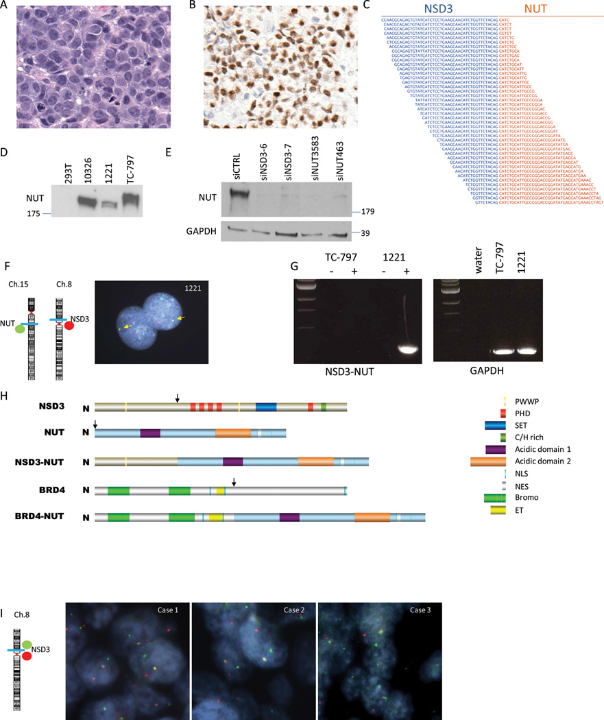
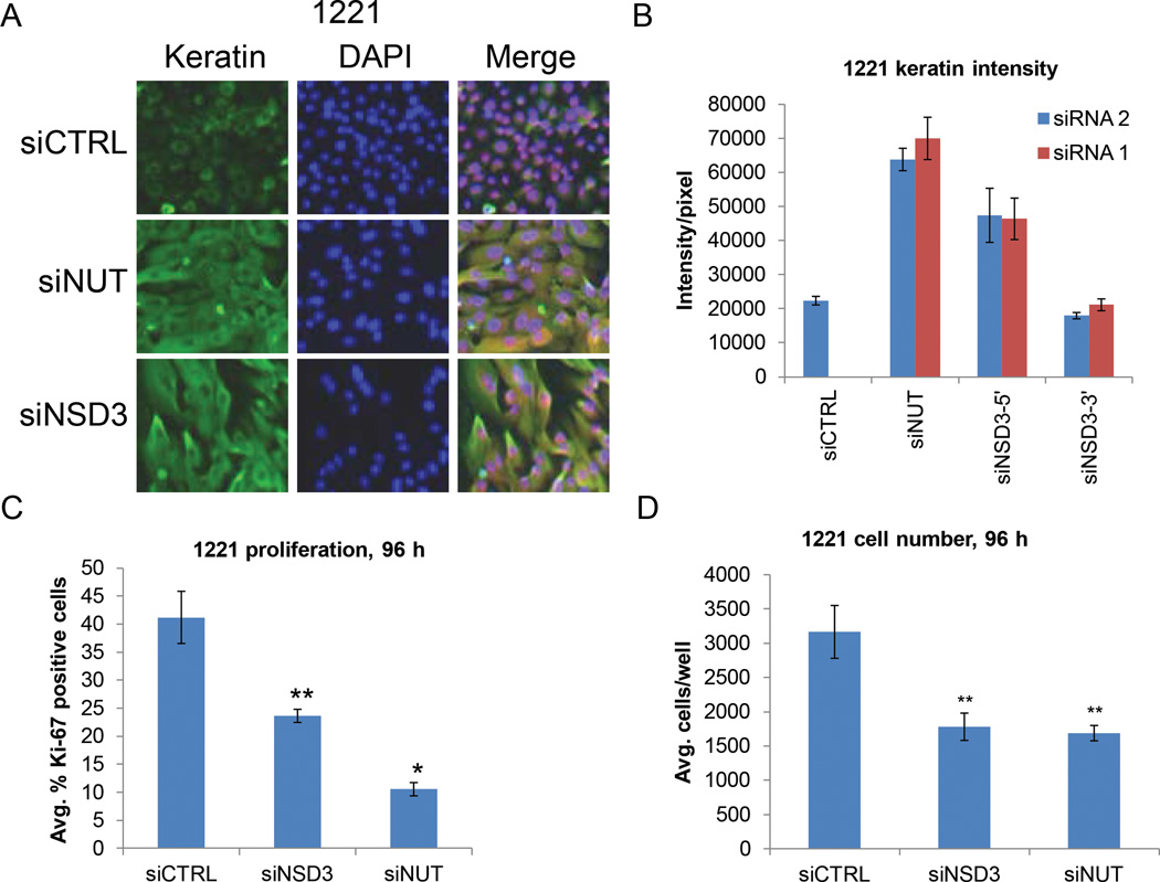
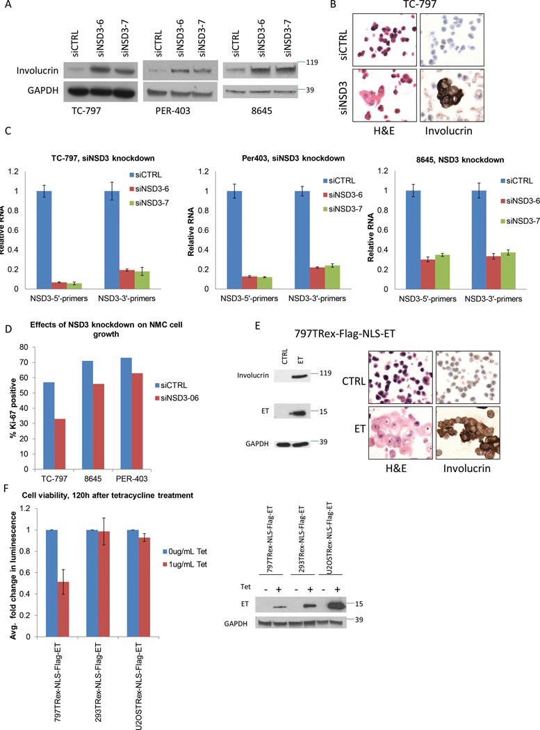
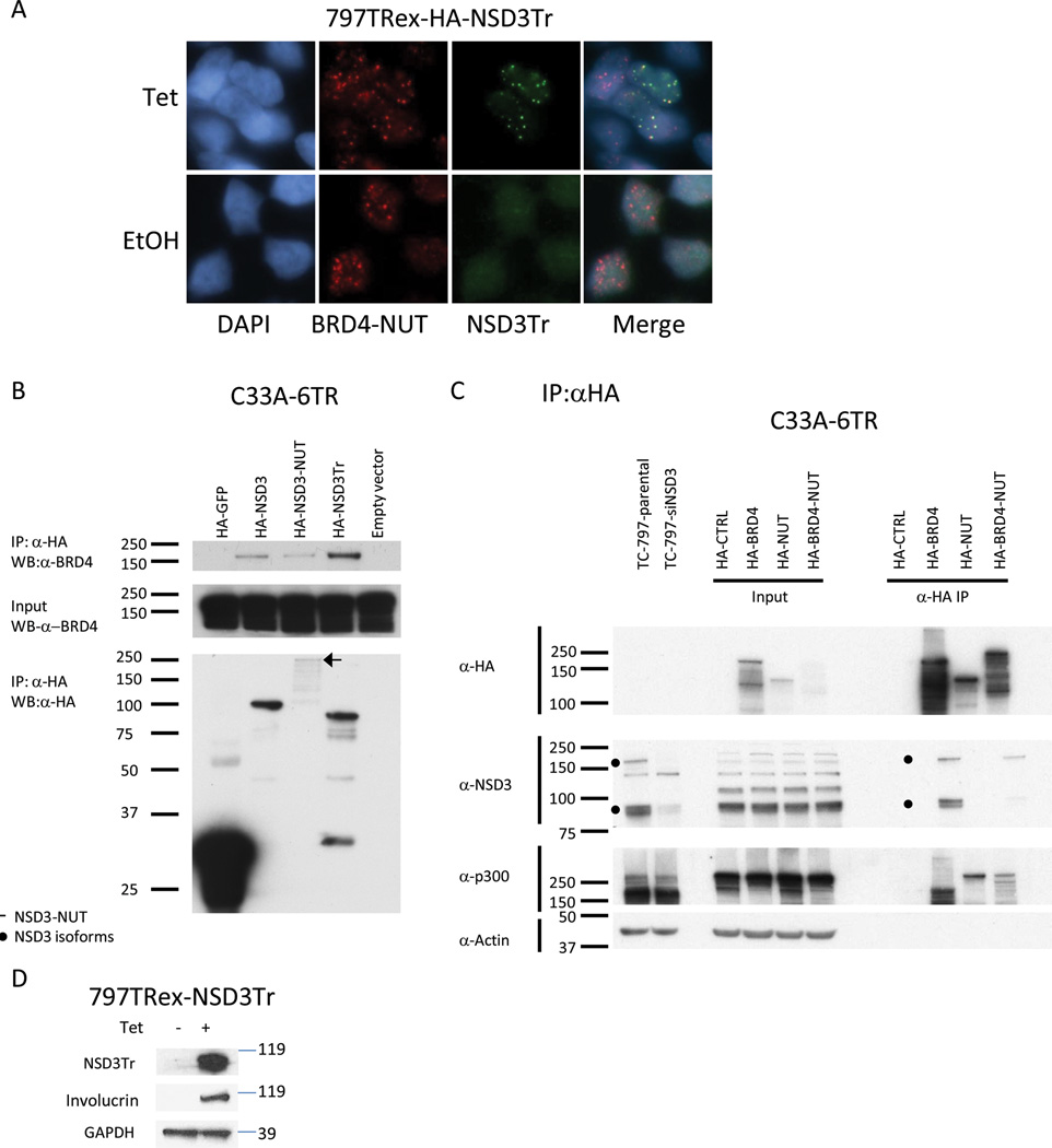
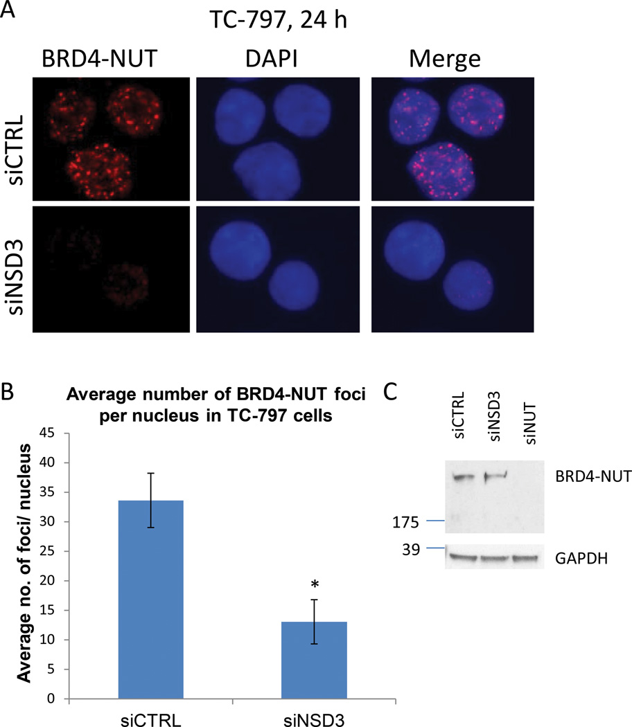
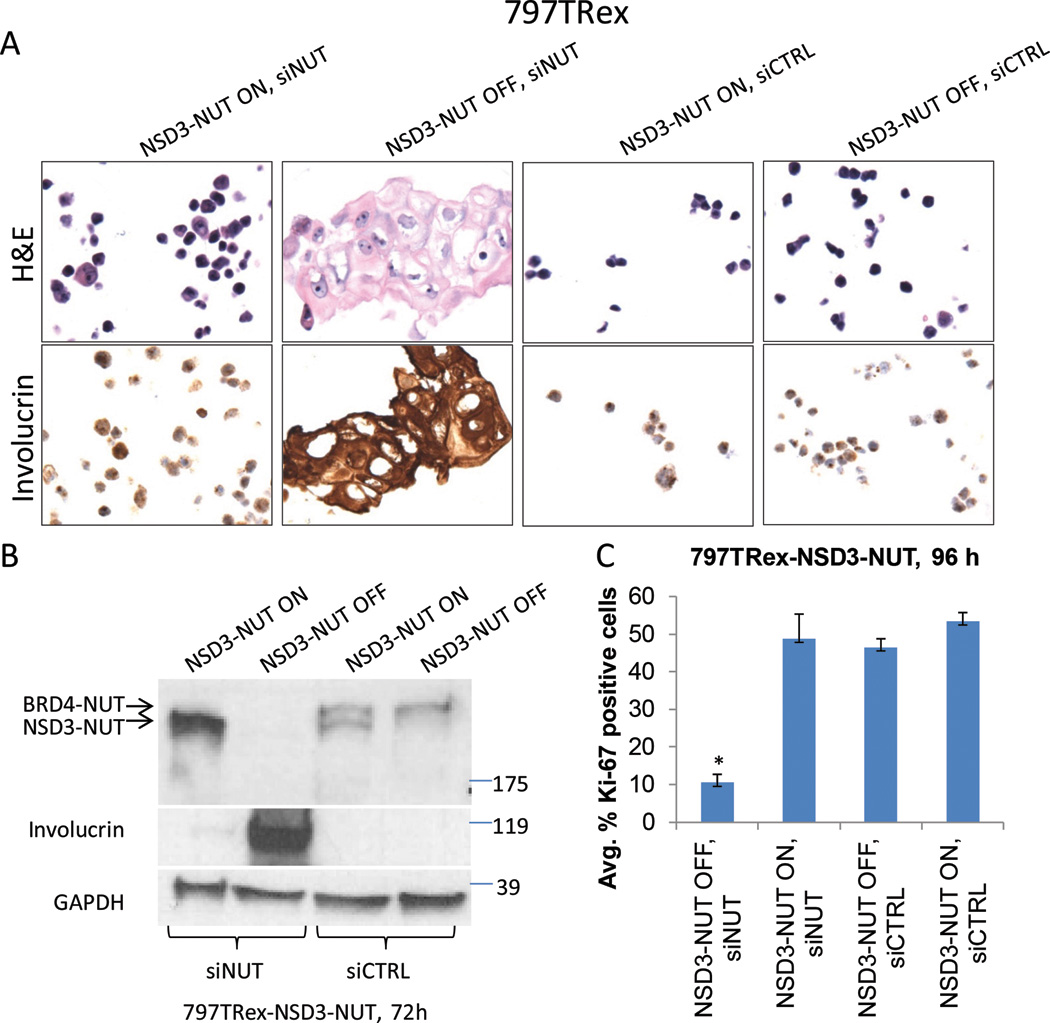
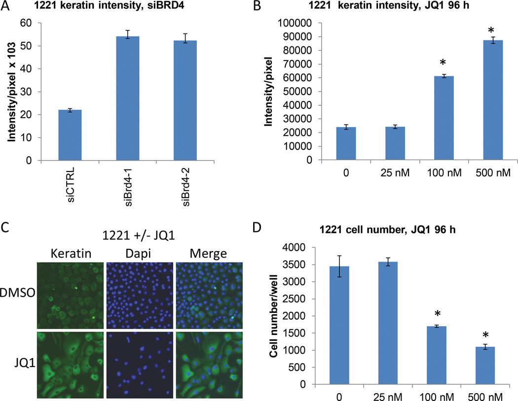
NUT midline carcinoma (NMC) is an aggressive subtype of squamous cell carcinoma that typically harbors BRD4/3-NUT fusion oncoproteins that block differentiation and maintain tumor growth. In 20% of cases, NUT is fused to uncharacterized non-BRD gene(s). We established a new patient-derived NMC cell line (1221) and demonstrated that it harbors a novel NSD3-NUT fusion oncogene. We find that NSD3-NUT is both necessary and sufficient for the blockade of differentiation and maintenance of proliferation in NMC cells. NSD3-NUT binds to BRD4, and BRD bromodomain inhibitors induce differentiation and arrest proliferation of 1221 cells. We find further that NSD3 is required for the blockade of differentiation in BRD4-NUT-expressing NMCs. These findings identify NSD3 as a novel critical oncogenic component and potential therapeutic target in NMC.

Significance: The existence of a family of fusion oncogenes in squamous cell carcinoma is unprecedented, and should lead to key insights into aberrant differentiation in NMC and possibly other squamous cell carcinomas. The involvement of the NSD3 methyltransferase as a component of the NUT fusion protein oncogenic complex identifies a new potential therapeutic target.


Conflict of interest statement

Disclosure of potential conflicts of interest: No potential conflicts of interest were disclosed.

Figures

Figure 1
Figure 1. A novel NSD3-NUT fusion is identified in NUT midline carcinoma
(A) Histology of the NMC from which the 1221 cell line was derived reveals a very poorly differentiated tumor (400× magnification). (B) Immunohistochemistry of the tumor using the anti-NUT monoclonal antibody, C52 (400× magnification). (C) RNA-sequencing reads spanning the junction of NSD3 and NUT. (D) Immunoblot of three NMC cell lines and 293T control cells stained with AX.1 polyclonal antibody to NUT. (E) Immunoblot of the 1221 cell line 48h following transfection with control (CTRL), NSD3, and NUT siRNAs stained with the AX.1 antibody to NUT. (F) NSD3-NUT dual color bring-together fluorescent in situ hybridization assay (1000× magnification) using BAC probes telomeric (3’) to NUT (green), and BAC probes centromeric (5’) to NSD3 (red) as depicted in the chromosomes 8 and 15 ideograms. Yellow arrows indicate NSD3-NUT fusions. (G) Gel electrophoresis of PCR of TC-797 and 1221 cell lines with (+) and without (−) reverse transcriptase reaction. (H) Schematic of the NSD3-NUT predicted encoded protein in comparison with NSD3, NUT, and BRD4-NUT. Abbreviations: PWWP, Pro-Trp-Trp-Pro motif; PHD, PHD finger (Plant Homeo Domain); SET, Su(var)3–9, Enhancer-of-zeste and Trithorax domain; C/H rich, Cys–His-rich region; NLS, nuclear localization sequence; NES, nuclear export signal sequence; Bromo, bromodomain; ET, extra-terminal domain. Arrows indicated breakpoints. (I) NSD3 dual color split-apart fluorescent in situ hybridization assay using BAC probes flanking NSD3, as depicted in the chromosome 8 ideogram, depicted in three NMCs designated cases 1–3. All photomicrographs are identical magnification (1000×).
Figure 2
Figure 2. NSD3-NUT is required for the blockade of differentiation and maintenance of proliferation in 1221 NMC cells
(A) High throughput 384 well plate immunofluorescent assay of keratin using the DAPI nuclear counterstain in 1221 cells 72 h following transfection with control, NUT, or NSD3 siRNAs. Representative photos are identical magnification (400X). (B) Using the high-throughput assay in (A) quantitative analysis of keratin intensity was compared in 1221 cells 72 h following transfection with control, NUT, NSD3-5’ (targets both NSD3-NUT and NSD3-full length), and NSD3-3’ (targets the NSD3 portion not included in NSD3-NUT). Two different siRNAs were used for each gene or region targeted. Representative results from one of three biological replicates, each performed in triplicate, are shown. Error bars indicate the mean ± SD of the triplicate wells. (C) Proliferation assay (Ki-67 fraction) using the high-throughput assay comparing 1221 cells transfected with control, NUT, and NSD3 siRNAs. Shown are averages of three biological replicates, each performed in triplicate. Error bars indicate the mean ± SD of the three biological replicates. (D) Cell number using the high-throughput assay comparing 1221 cells transfected with control, NUT, and NSD3 siRNAs. Shown are averages of three biological replicates, each performed in triplicate. Error bars indicate the mean ± SD of the three biological replicates. * p<0.01; **p < 0.05.
Figure 3
Figure 3. Wild type NSD3 is required for the blockade of differentiation in BRD4-NUT-expressing NMC cells
(A) Immunoblots of BRD4-NUT-positive NMC cell lines TC-797, PER-403, and 8645 120h following transfection with control and NSD3 siRNAs stained with the terminal squamous differentiation marker, involucrin, using GAPDH as loading control. (B) Representative photomicrographs of TC-797s 120 h following transfection with either control, or NSD3 siRNAs stained either with hematoxylin and eosin (H&E) for morphology, or involucrin immunohistochemistry. All photos are identical magnification (400×). (C) Quantitative RT-PCR of NSD3 levels 24 h following transfection of control or NSD3 siRNAs. Primers were either 5’ of the breakpoint (NSD3-5’ primers), or 3’ of the breakpoint (NSD3-3’ primers) with NUT. Results are of a single biological replicate performed in triplicate. Error bars indicate the mean ± SD of the triplicate wells. (D) Proliferation assay (Ki-67 fraction) comparing BRD4-NUT-positive TC-797, 8645, and PER-403 NMC cells transfected with control and NSD3-6 siRNAs. Three hundred cells were counted per cell block. (E) 797TRex cells induced to express FLAG-tagged NLS-ET domain of BRD4 for 120 h. Immunoblot was stained with anti-involucrin (Inv), anti-FLAG, or anti-GAPDH (left). Cell block preparations were H&E stained, or subjected to involucrin immunohistochemistry (right). All photos are identical magnification (400×). (F) Cell viability assay (CellTiter-Glo) of 797TRex, 293TRex, and U2OSTRex cells induced to express FLAG-tagged NLS-ET domain for 120h. Results are the average of three biological replicates, each performed in quadruplet and normalized to the negative control (ethanol vehicle control) for each cell line. Error bars indicate the mean ± SD of the three biological replicates. Immunoblot demonstrating NLS-FLAG-ET expression was stained with anti-FLAG, or anti-GAPDH (right).
Figure 4
Figure 4. The N-terminus of NSD3 associates with BRD4 and BRD4-NUT
(A) Immunofluorescence microscopy of 797TRex cells induced to express the HA-tagged portion of NSD3 included in NSD3-NUT (NSD3Tr) for 24 h stained with anti-NUT monoclonal antibody (red), and anti-HA monoclonal antibody (green). (B) Immunoblot of anti-HA immunoprecipitations of tet-repressor-positive C33A cell (C33A-6TR) lysates following induction of expression of HA-tagged NSD3 variants, HA-NSD3 (full length), HA-NSD3-NUT and HA-NSD3-tr (NSD3 portion of the NSD3-NUT fusion protein). Indicated proteins were detected using anti-HA, and anti-Brd4 antibodies. The smaller bands are degraded protein. (C) Immunoblot of anti-HA immunoprecipitations of C33A-6TR lysates following induction of expression of HA-tagged NUT, BRD4, and BRD4-NUT constructs stained with anti-HA, -NSD3, -p300, and -actin antibodies. To identify the NSD3-specific bands, lysates from TC-797s subjected to siRNA knockdown of NSD3 are shown. (D) Immunoblot of 797TRex lystes 120h following induction of expression of BioTAP-tagged NLS-fusion construct of NSD3Tr stained with anti-involucrin, -PAP (recognizes the protein A moiety of the BioTAP tag), and –GAPDH antibodies.
Figure 5
Figure 5. BRD4-NUT foci are dependent on NSD3
(A) Immunofluorescence microscopy of TC-797 cells 24 h following transfection with control or NSD3-6 siRNAs stained with monoclonal antibody to NUT. All photos are identical magnification (1000×). (B) Quantitation of BRD4-NUT foci was performed in triplicate and the averages of the three experiments. Error bars indicate the mean ± SD of triplicate experiments. *p < 0.005. (C) Immunoblot of TC-797 lysates 24 h following transfection with control, NSD3-6, or NUT siRNAs stained with anti-NUT polyclonal antibody, AX.1.
Figure 6
Figure 6. NSD3-NUT can replace the function of BRD4-NUT to block differentiation
(A) H&E and anti-involucrin immunohistochemistry micrographs of 797TRex cells with tetracycline (ON), or treated with vehicle (OFF) to express NSD3-NUT 120 h following transfection with either control or NUT 3’UTR siRNA. All photos are identical magnification (400×). (B) Immunoblots using lysates corresponding to the experiment in (A) were performed for the differentiation marker, involucrin, NSD3-NUT, and BRD4-NUT using antibodies to NUT. (C) Quantification of immunohistochemical Ki-67 proliferation fraction of 797TRex cells induced to express NSD3-NUT 120 h following transfection with either control or NUT 3’UTR siRNA as in (A). Results are the average of three biological replicates performed using the 384-well high throughput assay as in Figure 2A, each performed in triplicate. Error bars indicate the mean ± SD of the three biological replicates. *p <0.0001.
Figure 7
Figure 7. BRD4 inhibition arrests proliferation and induces differentiation of NSD3-NUT-expressing NMC cells
(A) Using the 384-well plate high-throughput assay exhibited in Figure 2A, quantitative analysis of keratin intensity was compared in 1221 cells 72 h following transfection with control versus BRD4 siRNAs. Representative results from one of three biological replicates, each performed in triplicate, are shown. Error bars indicate the mean ± SD of triplicate wells. (B) Using the high-throughput assay (above), quantitative analysis of keratin intensity was compared in 1221 cells 72 h following treatment with a dose range of JQ1 versus DMSO vehicle control. Results are the average of three biological replicates performed using the 384-well high throughput assay, each performed in triplicate. Error bars indicate the mean ± SD of the three biological replicates.* p <0.01 (C) Representative immunofluorescence microscopy of 1221 cells treated as in (B), with vehicle control or 500nM JQ1 for 72h. All photos are identical magnification (400×). (D) Cell number using the high-throughput assay comparing 1221 cells 72 h following treatment with increasing concentrations of JQ1 versus DMSO vehicle control. Results are the average of three biological replicates, each performed in triplicate. Error bars indicate the mean ± SD of the three biological replicates.* p <0.01

References

    1. French CA, Miyoshi I, Kubonishi I, Grier HE, Perez-Atayde AR, Fletcher JA. BRD4-NUT fusion oncogene: a novel mechanism in aggressive carcinoma. Cancer research. 2003;63(2):304–307. - PubMed
    1. French CA, Ramirez CL, Kolmakova J, Hickman TT, Cameron MJ, Thyne ME, et al. BRD-NUT oncoproteins: a family of closely related nuclear proteins that block epithelial differentiation and maintain the growth of carcinoma cells. Oncogene. 2008;27(15):2237–2242. Epub 2007/10/16. - PubMed
    1. Bauer DE, Mitchell CM, Strait KM, Lathan CS, Stelow EB, Luer SC, et al. Clinicopathologic Features and Long-Term Outcomes of NUT Midline Carcinoma. Clinical cancer research : an official journal of the American Association for Cancer Research. 2012;18(20):5773–5779. Epub 2012/08/17. - PMC - PubMed
    1. Filippakopoulos P, Qi J, Picaud S, Shen Y, Smith WB, Fedorov O, et al. Selective inhibition of BET bromodomains. Nature. 2010;468(7327):1067–1073. Epub 2010/09/28. - PMC - PubMed
    1. Matzuk MM, McKeown MR, Filippakopoulos P, Li Q, Ma L, Agno JE, et al. Small-molecule inhibition of BRDT for male contraception. Cell. 2012;150(4):673–684. Epub 2012/08/21. - PMC - PubMed

Publication types

MeSH terms