. 2014 May 29;10(5):e1004403.
doi: 10.1371/journal.pgen.1004403. eCollection 2014.

Rapid evolution of PARP genes suggests a broad role for ADP-ribosylation in host-virus conflicts

Affiliations

Rapid evolution of PARP genes suggests a broad role for ADP-ribosylation in host-virus conflicts

Matthew D Daugherty et al. PLoS Genet. .

Abstract

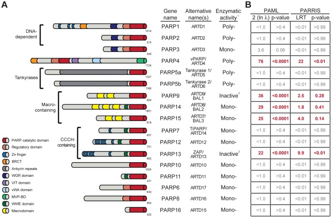
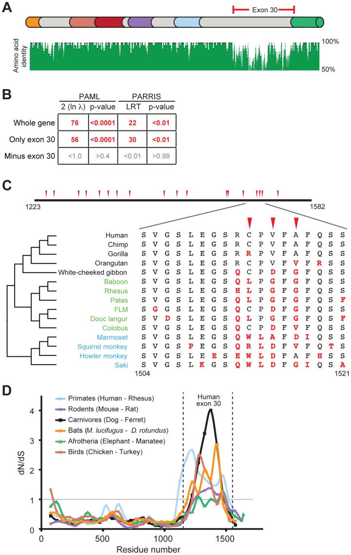
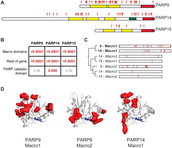
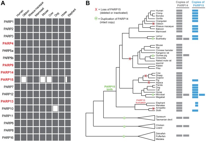
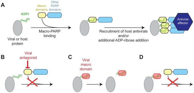
Post-translational protein modifications such as phosphorylation and ubiquitinylation are common molecular targets of conflict between viruses and their hosts. However, the role of other post-translational modifications, such as ADP-ribosylation, in host-virus interactions is less well characterized. ADP-ribosylation is carried out by proteins encoded by the PARP (also called ARTD) gene family. The majority of the 17 human PARP genes are poorly characterized. However, one PARP protein, PARP13/ZAP, has broad antiviral activity and has evolved under positive (diversifying) selection in primates. Such evolution is typical of domains that are locked in antagonistic 'arms races' with viral factors. To identify additional PARP genes that may be involved in host-virus interactions, we performed evolutionary analyses on all primate PARP genes to search for signatures of rapid evolution. Contrary to expectations that most PARP genes are involved in 'housekeeping' functions, we found that nearly one-third of PARP genes are evolving under strong recurrent positive selection. We identified a >300 amino acid disordered region of PARP4, a component of cytoplasmic vault structures, to be rapidly evolving in several mammalian lineages, suggesting this region serves as an important host-pathogen specificity interface. We also found positive selection of PARP9, 14 and 15, the only three human genes that contain both PARP domains and macrodomains. Macrodomains uniquely recognize, and in some cases can reverse, protein mono-ADP-ribosylation, and we observed strong signatures of recurrent positive selection throughout the macro-PARP macrodomains. Furthermore, PARP14 and PARP15 have undergone repeated rounds of gene birth and loss during vertebrate evolution, consistent with recurrent gene innovation. Together with previous studies that implicated several PARPs in immunity, as well as those that demonstrated a role for virally encoded macrodomains in host immune evasion, our evolutionary analyses suggest that addition, recognition and removal of ADP-ribosylation is a critical, underappreciated currency in host-virus conflicts.


Conflict of interest statement

The authors have declared that no competing interests exist.

Figures

Figure 1
Figure 1. Several PARP genes are evolving under positive selection.
(A) Schematic domain structures of human PARP proteins are shown (not to scale). Numbers to the bottom right of the protein schematic indicate the total length, in amino acids, of each protein. BRCT: BRCA1 C-terminus domain; VIT: vault inter-trypsin domain; vWA: von Willebrand factor type A; MVP-BD: major vault protein binding domain. 1PARPs are categorized as either poly-ADP-ribosyltransferases (Poly-), mono-ADP-ribosyltransferases (Mono-) or inactive based on presence of conserved motifs and, when available, data using enzymatic assays , . 2PARP9 and PARP13 lack one or more catalytic residues conserved in all other PARPs and are therefore predicted to lack catalytic activity, although it is unknown whether they still bind ADP-ribose or ADP-ribosylated proteins . (B) Results of maximum likelihood tests for positive selection. The five PARP genes showing strong signatures of positive selection are shown in red. Shown at left are values derived from PAML indicating twice the log difference between an evolutionary model that allows positive selection (M8) versus a model that disallows position selection (M8a). P-values indicate whether the model that allows positive selection better fits the data. PARRIS-derived LRT (likelihood ratio test) values also report the difference between the log-likelihood values for a model allowing positive selection versus a model that does not. Using seven orthologs in PARRIS only gave a statistically significant p-value (<0.01) for PARP4 and PARP13, however analysis of additional sequences of PARP9, 14 and 15 found that these genes also met statistical significance (see Results). Additional analyses are reported in Figure S1.
Figure 2
Figure 2. Positive selection in primate PARP4 is localized to exon 30.
(A) The domain structure of human PARP4 protein is shown with domain colors as in Figure 1A. The region of the protein corresponding to human exon 30 is bracketed in red. Also shown below the schematic is an amino acid identity plot calculated using seven publicly available primate PARP4 sequences. (B) Results of maximum likelihood tests for positive selection from seven publically available PARP4 sequences using the entire gene, exon 30 alone or the entire gene except exon 30. Columns are as in Figure 1B. (C) An expanded view of exon 30 (residues 1223 to 1582 in human PARP4). Codons evolving under recurrent positive selection in an analysis of 15 primate species are marked with red triangles (posterior probabilities greater than 0.95) (see also Table S4). Below is a further expanded view of the alignment of residues 1504 to 1521 across all primates, with the phylogenetic tree shown to the left (Hominoids (black), Old World monkeys (green) and New World monkeys (blue)). Residues in red are changes from the human protein. (D) A sliding window dN/dS analysis of several pairs of vertebrate PARP4 genes using a window size of 150 codons and a step size of 50 codons. The grey horizontal line marks a dN/dS value of 1, indicating neutral evolution. The vertical dashed lines outline the relative position of human exon 30. dN/dS values and confidence intervals from analyses of the largest exon of PARP4 from various vertebrate pairwise comparisons are shown in Table S3.
Figure 3
Figure 3. Widespread distribution of positive selection in macro-PARP genes.
(A) The domain structure of each macro-PARP gene is shown with domain colors as in Figure 1A. Codons evolving under recurrent positive selection are marked with red triangles as in Figure 2 (see also Tables S6-S8). (B) P–values derived from PAML tests of positive selection on all sequenced macro-PARP orthologs using either macrodomains alone, the entire gene minus the macrodomains or just the catalytic domain. Values in red indicate strong evidence of positive selection, while those in grey indicate a lack of statistically significant evidence for positive selection. (C) Phylogenetic tree of individual macrodomains from macro-PARPs, rooted using human MacroD2 as an outgroup. Macrodomains in italics lack a C-terminal extension found in most macrodomains and lack one or more of the putative catalytic motifs required for ADP-ribosylhydrolase activity (Figure S5). This suggests these macrodomains may be able to recognize, but are unlikely to catalyze removal of, ADP-ribosylation. To the right is a schematic of each macrodomain with positively selected residues indicated in red (posterior probabilities greater than 0.95) (see also Alignment S3). (D) Location of positively selected sites in PARP macrodomains mapped on to the structure of the first macrodomain from PARP14 in complex with ADP-ribose (PDB code: 3Q6Z). ADP-ribose is shown as blue sticks. Residues shown as red surfaces are those that have evolved under positive selection in the indicated macrodomain.
Figure 4
Figure 4. Recurrent gain and loss of PARP14 and PARP15.
(A) Intact PARP genes were identified in representative (high quality assembly) mammalian genomes. Filled rectangles indicate the presence of an intact gene, open rectangles indicate a gene loss. The half-filled rectangle indicates a large N-terminal truncation of PARP15 in the elephant genome. (B) Intact PARP14 and 15 homologs were compiled from publicly available vertebrate genomes (see Figure S6). A phylogenetic tree of queried species is shown with PARP14 duplications (including the birth of PARP15) and losses of PARP15 indicated. To the right is an indication of the number of intact PARP14 and PARP15 homologs in each genome, as indicated by the filled rectangles.
Figure 5
Figure 5. Model for genetic conflict involving PARP macrodomains.
(A) Model for macro-PARP function. ADP-ribosylated host or viral proteins may be a signal for recruitment of PARP9, 14 or 15, which could facilitate additional recruitment of antiviral effectors, and amplify the initial ADPr signal. (B–D) Three models for how viruses may antagonize macro-PARP function. Viruses lacking their own macrodomains may use other proteins to directly antagonize macro-PARP proteins (B), driving recurrent positive selection in macro-PARP genes to escape antagonism. Macrodomains encoded by viruses (e.g. corona- and togaviruses) may catalyze the removal of ADPr (C) or compete with macro-PARPs for binding to ADPr (D) in order to antagonize host ADPr-mediated signaling.

References

    1. Salomon D, Orth K (2013) What pathogens have taught us about posttranslational modifications. Cell Host Microbe 14: 269–279. - PMC - PubMed
    1. Adhya D, Basu A (2010) Epigenetic modulation of host: new insights into immune evasion by viruses. J Biosci 35: 647–663. - PubMed
    1. Isaacson MK, Ploegh HL (2009) Ubiquitination, ubiquitin-like modifiers, and deubiquitination in viral infection. Cell Host Microbe 5: 559–570. - PMC - PubMed
    1. Hottiger MO, Hassa PO, Luscher B, Schuler H, Koch-Nolte F (2010) Toward a unified nomenclature for mammalian ADP-ribosyltransferases. Trends Biochem Sci 35: 208–219. - PubMed
    1. Ziegler M (2000) New functions of a long-known molecule. Emerging roles of NAD in cellular signaling. Eur J Biochem 267: 1550–1564. - PubMed

Publication types

MeSH terms