

Laparoscopic resection of a retroperitoneal pelvic schwannoma
- PMID: 24876325
- PMCID: PMC3895046
- DOI: 10.1093/jscr/rjt122
Laparoscopic resection of a retroperitoneal pelvic schwannoma
Abstract
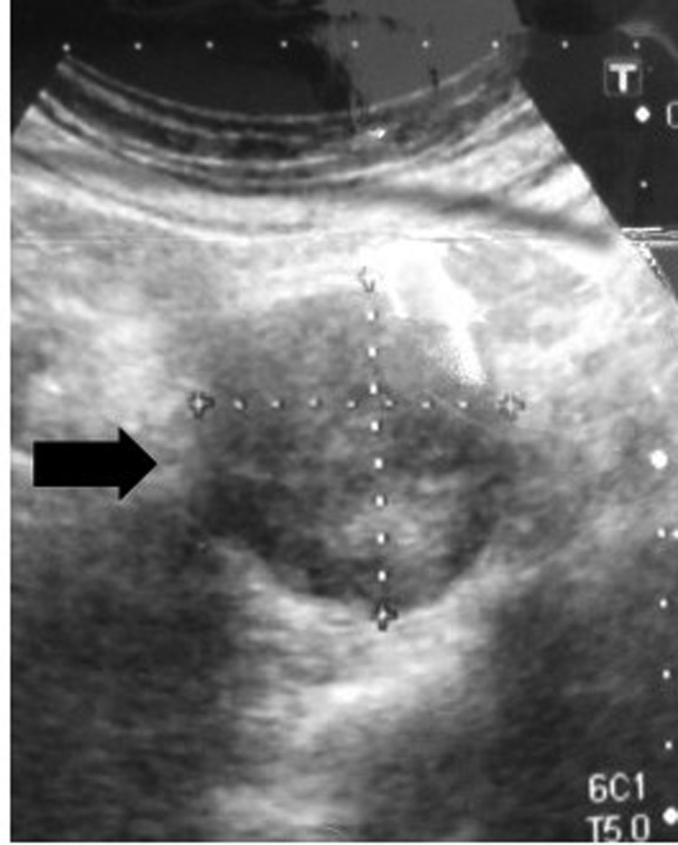
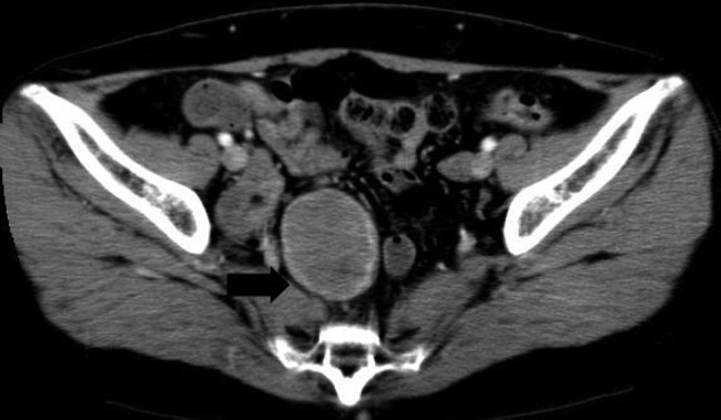
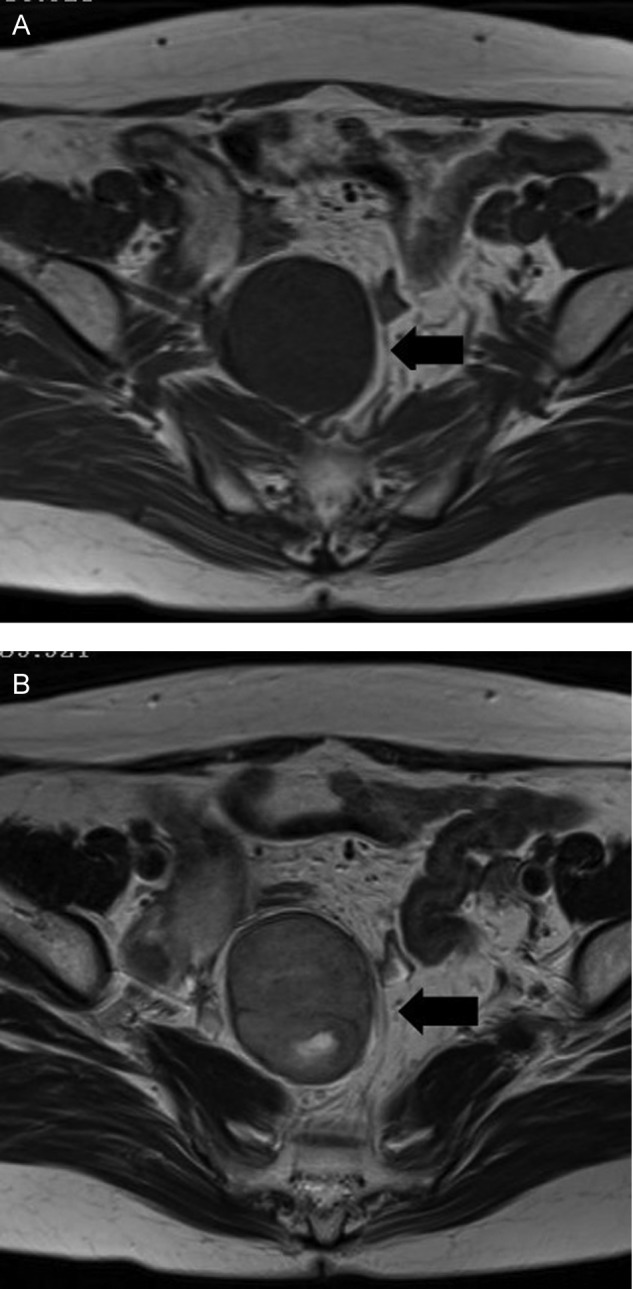
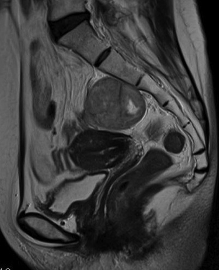
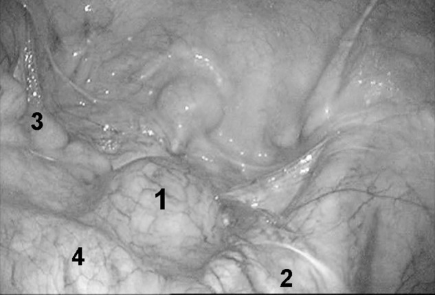
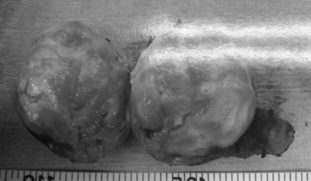
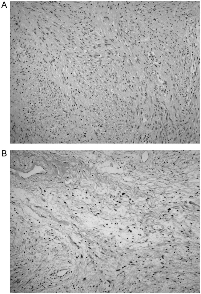
Schwannomas are rarely located in the pelvis. A 54-year-old woman was found incidentally to have a tumor in the abdomen. Abdominal computed tomography and magnetic resonance imaging revealed a well-defined, heterogeneous tumor, 5 cm in diameter, in the pelvic cavity. With a diagnosis of a mesenteric tumor, a laparoscopic procedure was performed. Intra-operatively, an elastic tumor was identified in the pelvis adjacent to the right internal iliac vein and ureter. The tumor was dissected free from adjacent structures using Liga-Sure and blunt maneuvers. A complete laparoscopic excision was performed. Histopathological examination revealed a benign schwannoma. The patient had an uneventful post-operative course, and was discharged on the fourth post-operative day. Laparoscopic treatment is useful and feasible for retroperitoneal pelvic schwannoma, with minimal invasiveness and an early post-operative recovery. Thus, this procedure may be the first-choice surgical procedure for retroperitoneal pelvic schwannomas.
Published by Oxford University Press and JSCR Publishing Ltd. All rights reserved. © The Author 2014.
Figures
Similar articles
-
Laparoscopic excision of a retroperitoneal schwannoma: A case report.Asian J Endosc Surg. 2019 Apr;12(2):192-196. doi: 10.1111/ases.12607. Epub 2018 May 28. Asian J Endosc Surg. 2019. PMID: 29808539
-
Laparoscopic Extirpation of a Schwannoma in the Lateral Pelvic Space.Case Rep Surg. 2016;2016:1351282. doi: 10.1155/2016/1351282. Epub 2016 Nov 9. Case Rep Surg. 2016. PMID: 27900226 Free PMC article.
-
Laparoscopic Resection of An Extragastrointestinal Stromal Tumor in the Presacral Area.J Minim Invasive Gynecol. 2019 Jul-Aug;26(5):812-813. doi: 10.1016/j.jmig.2018.10.023. Epub 2018 Nov 2. J Minim Invasive Gynecol. 2019. PMID: 30395935
-
[A case of retroperitoneal schwannoma treated by laparoscopic resection].Hinyokika Kiyo. 2009 Mar;55(3):129-31. Hinyokika Kiyo. 2009. PMID: 19378822 Review. Japanese.
-
Hybrid open/closed resection procedure for ancient retroperitoneal Schwannoma: case report and review of the literature.Acta Chir Belg. 2016 Oct;116(5):289-292. doi: 10.1080/00015458.2016.1177999. Epub 2016 Jun 2. Acta Chir Belg. 2016. PMID: 27426667 Review.
Cited by
-
A giant ancient schwannoma mimicking an adnexal mass: Case report.Medicine (Baltimore). 2016 Jul;95(30):e4240. doi: 10.1097/MD.0000000000004240. Medicine (Baltimore). 2016. PMID: 27472696 Free PMC article.
-
Clinical characteristics and prognosis of cystic degeneration in retroperitoneal schwannoma: A retrospective study of 79 patients.Cancer Med. 2023 Mar;12(5):5615-5629. doi: 10.1002/cam4.5411. Epub 2022 Nov 28. Cancer Med. 2023. PMID: 36440500 Free PMC article.
-
Laparoscopic resection of a retroperitoneal schwannoma located in the hepatic hilus.Surg Case Rep. 2015 Dec;1(1):18. doi: 10.1186/s40792-015-0024-6. Epub 2015 Feb 19. Surg Case Rep. 2015. PMID: 26943386 Free PMC article.
-
Retroperitoneal Ancient Schwannoma: A Case Report.Rev Urol. 2015;17(3):190-3. Rev Urol. 2015. PMID: 26543435 Free PMC article. Review.
-
Clinical Characteristics and Treatment Strategy of Retroperitoneal Schwannoma Adjacent to Important Abdominal Vessels: Three Case Reports and Literature Review.Front Surg. 2021 Jan 14;7:605867. doi: 10.3389/fsurg.2020.605867. eCollection 2020. Front Surg. 2021. PMID: 33585546 Free PMC article.
References
-
- Borghese M, Corigliano N, Gabriele R, Antoniozzi A, Izzo L, Barbaro M, et al. Benign schwannoma of the pelvic retroperitoneum. Report of a case and review of the literature. G Chir. 2000;21:232–8. - PubMed
-
- Aubert J, Debiais F, Irani J, Dore B, Levillain P. Schwannome et appareil urinaire. A propos D`une tumeur du nerf obturateur [in French] Prog Urol. 1999;9:528–33. - PubMed
-
- Ueda M, Okamoto Y, Ueki M. A pelvic retroperitoneal schwannoma arising in the right paracolpium. Gynecol Oncol. 1996;60:480–3. - PubMed
-
- Tong RSK, Collier N, Kaya AH. Chronic sciatica secondary to retroperitoneal pelvic schwannoma. J Clin Neurosci. 2003;10:108–11. - PubMed
-
- Deboudt C, Labat J, Riant T, Bouchot O, Robert R, Rigaud J. Pelvic schwannoma: robotic laparoscopic resection. Neurosurgry. 2013;72:2–5. - PubMed
Publication types
LinkOut - more resources
Full Text Sources
Other Literature Sources
