. 2014 Jun;6(2):024113.
doi: 10.1088/1758-5082/6/2/024113. Epub 2014 May 30.

Bioreactor for modulation of cardiac microtissue phenotype by combined static stretch and electrical stimulation

Affiliations

Bioreactor for modulation of cardiac microtissue phenotype by combined static stretch and electrical stimulation

Jason W Miklas et al. Biofabrication. 2014 Jun.

Abstract

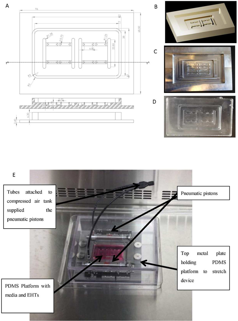
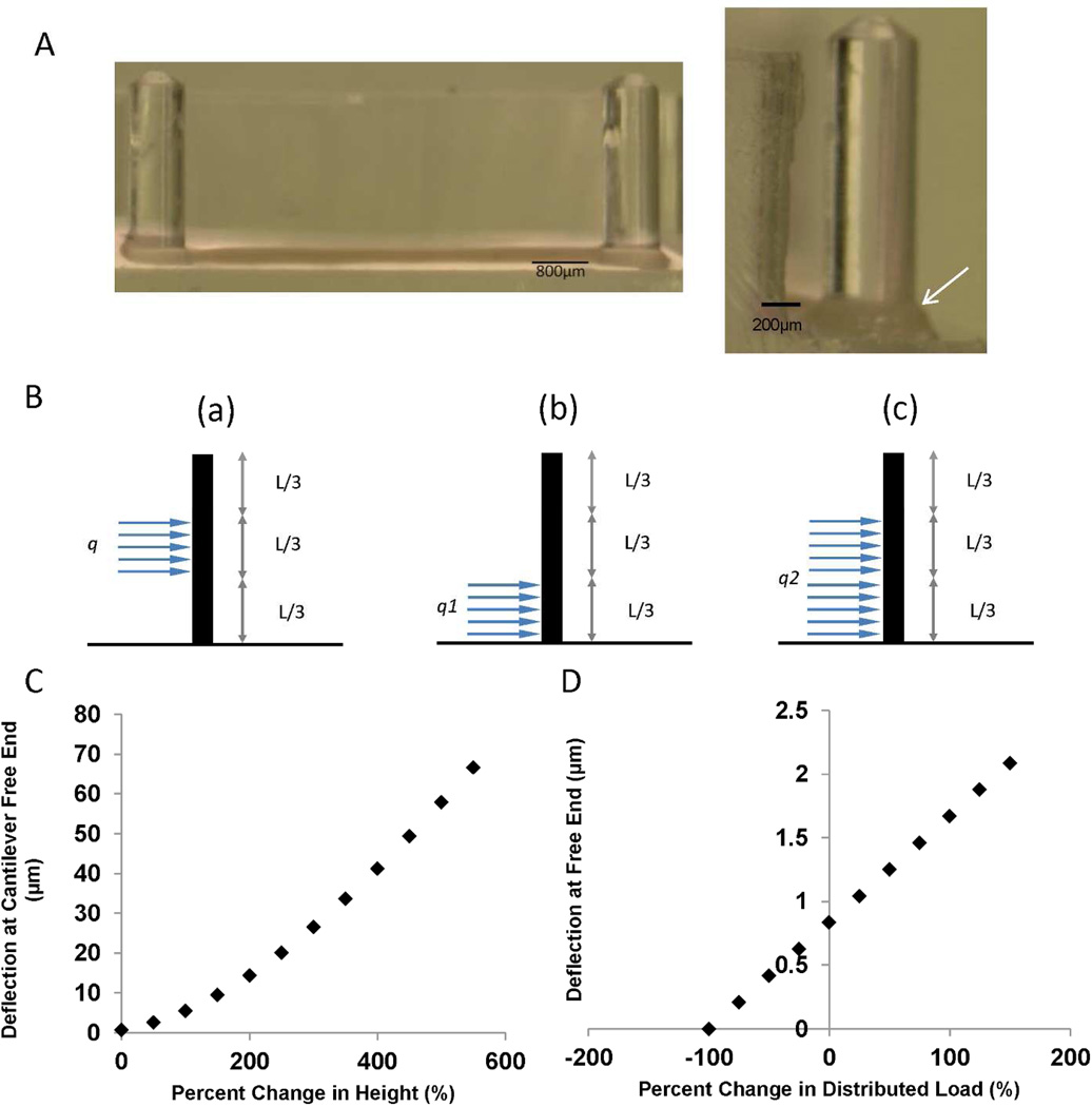
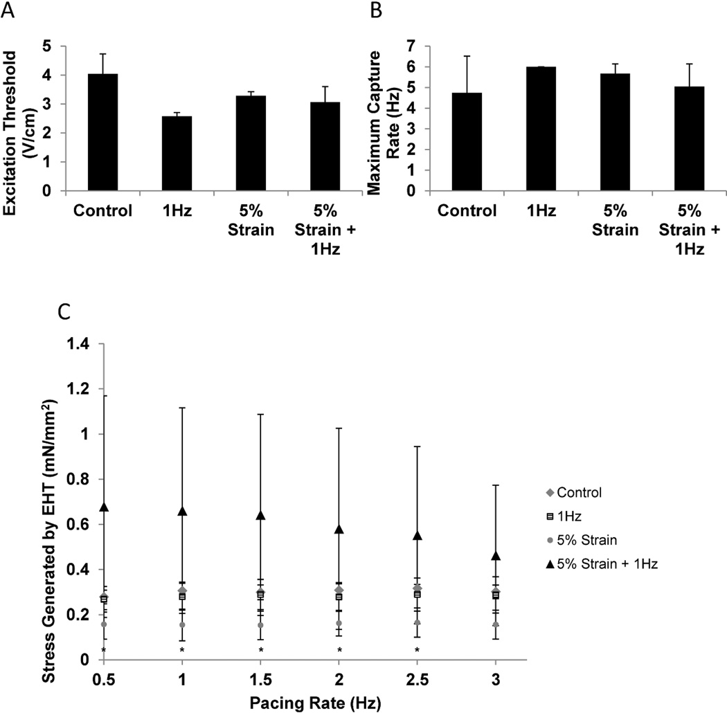
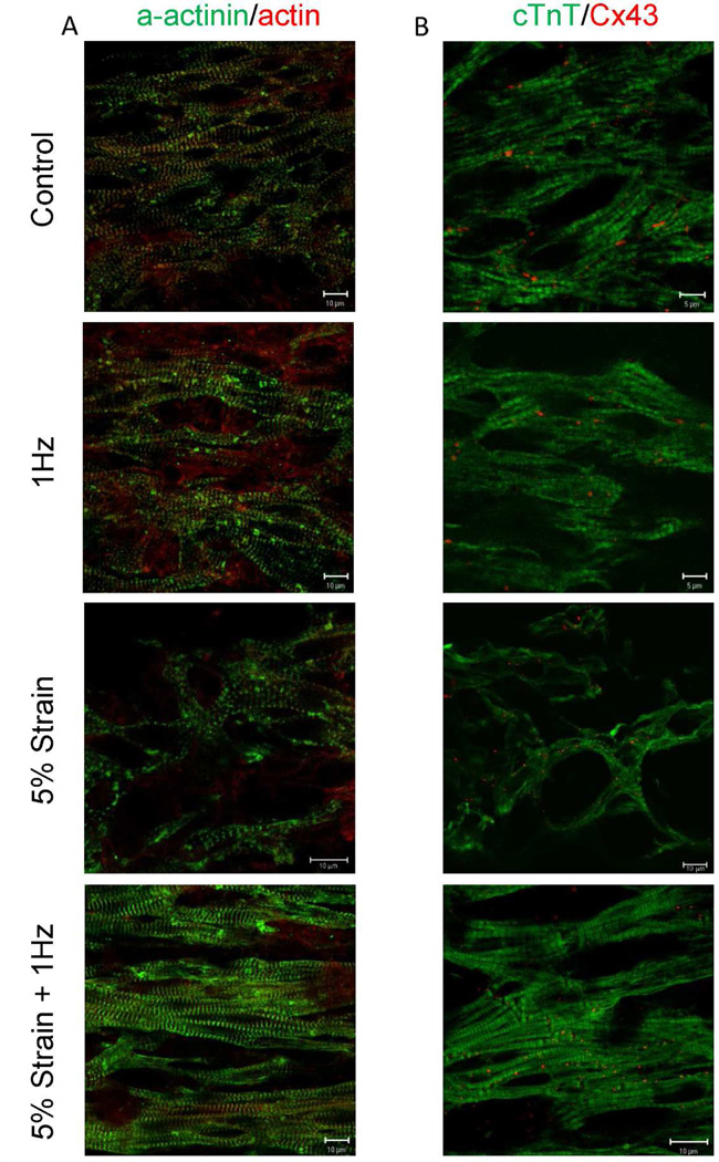
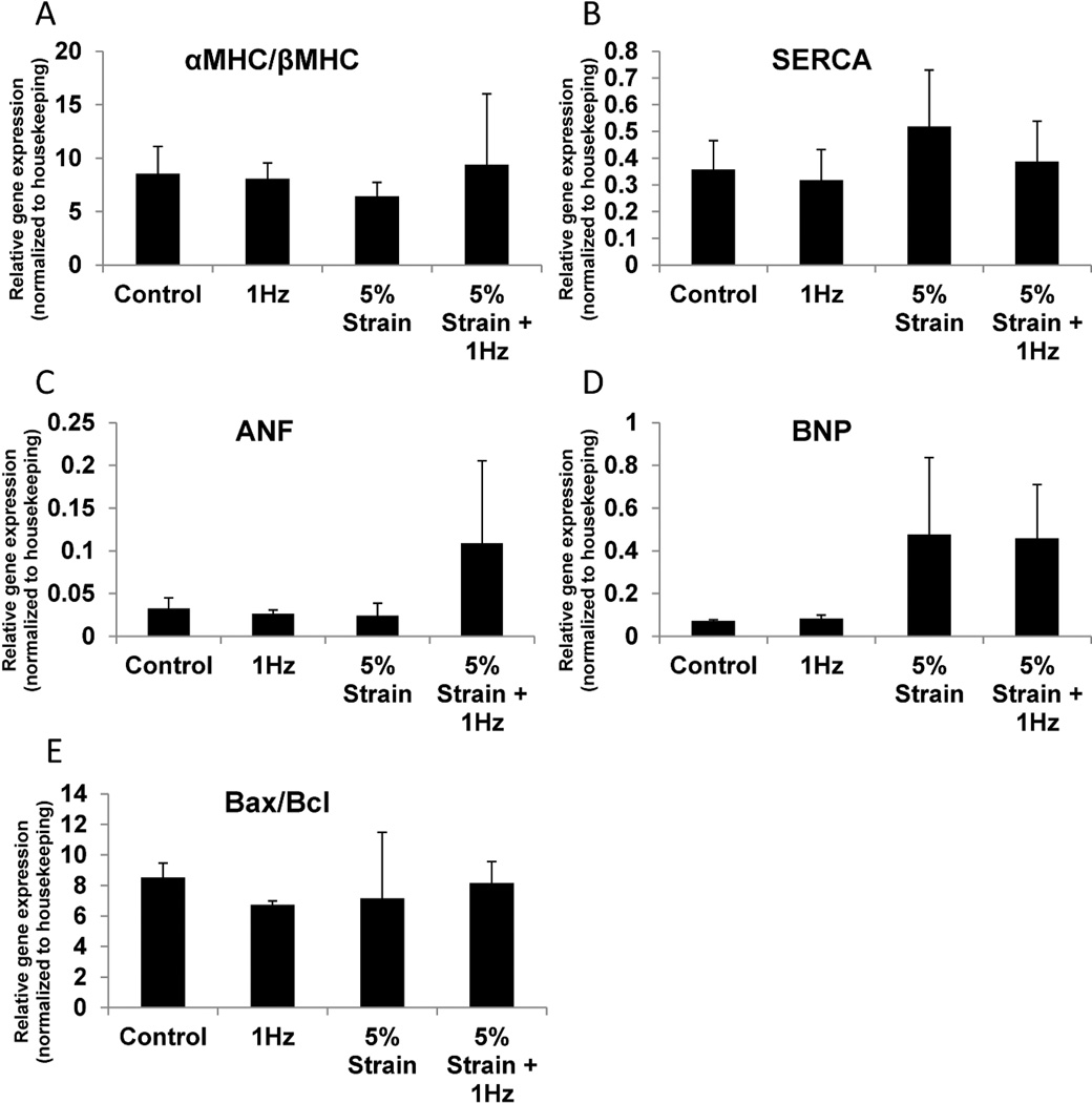
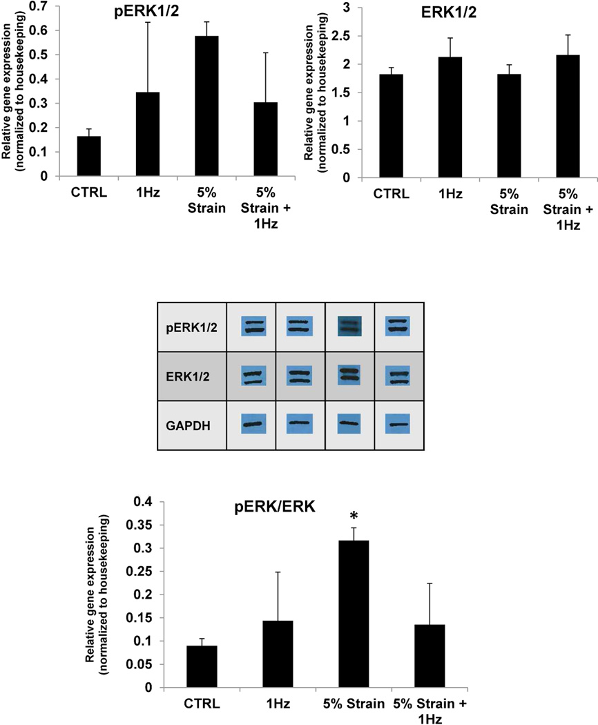
We describe here a bioreactor capable of applying electrical field stimulation in conjunction with static strain and on-line force of contraction measurements. It consisted of a polydimethylsiloxane (PDMS) tissue chamber and a pneumatically driven stretch platform. The chamber contained eight tissue microwells (8.05 mm in length and 2.5 mm in width) with a pair of posts (2.78 mm in height and 0.8 mm in diameter) in each well to serve as fixation points and for measurements of contraction force. Carbon rods, stimulating electrodes, were placed into the PDMS chamber such that one pair stimulated four microwells. For feasibility studies, neonatal rat cardiomyocytes were seeded in collagen gels into the microwells. Following 3 days of gel compaction, electrical field stimulation at 3-4 V cm(-1) and 1 Hz, mechanical stimulation of 5% static strain or electromechanical stimulation (field stimulation at 3-4 V cm(-1), 1 Hz and 5% static strain) were applied for 3 days. Cardiac microtissues subjected to electromechanical stimulation exhibited elevated amplitude of contraction and improved sarcomere structure as evidenced by sarcomeric α-actinin, actin and troponin T staining compared to microtissues subjected to electrical or mechanical stimulation alone or non-stimulated controls. The expression of atrial natriuretic factor and brain natriuretic peptide was also elevated in the electromechanically stimulated group.


Figures

Figure 2
Figure 2. Bioreactor platform for combined electrical field and static stretch stimulation
A) CAD drawing of a bioreactor well capable of housing four cardiac microtissues and two pairs of stimulating electrodes. Each cardiac tissue microwell contains a par of posts to be used for monitoring of tissue contractions. B) Three-dimensional CAD rendering of the bioreactor well used in the milling machine for production of aluminum mold. C) Aluminum mold produced by the milling process. D) PDMS bioreactor wells produced using aluminum mold. E) PDMS bioreactor well placed in the pneumatically driven stretch device capable of providing static stretch.
Figure 2
Figure 2. Validation of the strains and force of contraction measurements in the PDMS well of the bioreactor platform
A) Each cardiac tissue microwell contains a pair of posts that deflects as the tissue contracts. The tissue is generated by gel compaction of cardiomyocytes in a collagen gel (arrow). B) To calculate force of contraction, a beam deflection analytical model can be used to correlate imaged deflection of the post during a contraction cycle to force of contraction. If the tissue is not situated at the bottom of the post, a methods of superposition can be implemented to determine the distributed load on the post, where centrally positioned load (a) is modelled as a combination of two loads (b) and (c). The images are redrawn based on a schematic presented in reference [20] C) Sensitivity analysis of post free end deflection with varying tissue height along the post. A tissue of 0.4 mm thickness situated at the bottom of the post with an average point force of contraction of 0.2 mN was the base scenario (0% height change) in this graph. The tissue was then moved up the post and the deflection at the free end was calculated again based on the same force of contraction. D) Sensitivity analysis of post free end deflection with varying distributed load. A tissue of 0.4 mm thickness situated at the bottom of the post with an average point force of contraction of 0.2 mN (0.5 N/m) was the base scenario at 0% change in distributed load. The distributed load was then varied while assuming the tissue remained at the bottom of the post.
Figure 3
Figure 3. Functional properties of cardiac microtissues
A) Excitation threshold determined at the end of cultivation as a minimum voltage required to induce synchronous contraction. B) Maximum capture rate determined at the end of cultivation as the maximum tissue beating frequency. C) Force of contraction. Control- cardiac microtissues cultivated in the PDMS wells without electrical or mechanical stimulation. 1Hz- cardiac microtissues cultivated in the PDMS wells in the presence of electrical field stimulation at 1Hz. 5%- cardiac mictotissues cultivated in the PDMS stretched at 5% static strain without electrical stimulation. 5% strain + 1Hz- cardiac microtissues stretched at 5% static strain and concurrently subjected to electrical field stimulation at 1Hz. * denotes statistical significant by Two-way ANOVA between 5% and 5% + 1Hz groups at specific pacing frequencies. Data represented as average ± standard deviation, N=3.
Figure 4
Figure 4. Immunostaining of cardiac microtissues for sarcomeric and gap junctional proteins
A) Double staining for sarcomeric α-actinin (green) and actin red. B) Double staining for cardiac troponin T (green) and connexion-43 red. Control-cardiac microtissues cultivated in the PDMS wells without electrical or mechanical stimulation. 1Hz- cardiac microtissues cultivated in the PDMS wells in the presence of electrical field stimulation at 1Hz. 5% strain + 1Hz- cardiac microtissues stretched at 5% static strain and concurrently subjected to electrical field stimulation at 1Hz.
Figure 5
Figure 5. Quantitative polymerase chain reaction analysis cardiac genes in cardiac microtissues
A) Ratio of α-myosin heavy chain (MHC) to β-myosin heavy chain, B) Sarco/endoplasmic reticulum Ca2+ATP-ase (SERCA), C) Atrial natriuretic factor (ANF), D) Brain natriuretic peptide (BNP), E) Ratio of pro-apoptotic Bax to anti-apoptotic Bcl-2. Data represented as averages ± standard deviation, N=3.
Figure 6
Figure 6. Western blotting for extracellular signal regulated kinases (ERK)1/2 expression and phosphorylation
* denotes statistical significance (P=0.032) between 5% strain and control group using One-way ANOVA. Data represented as averages ± standard deviation, N=3.

References

    1. Yang L, Soonpaa MH, Adler ED, Roepke TK, Kattman SJ, Kennedy M, Henckaerts E, Bonham K, Abbott GW, Linden RM, Field LJ, Keller GM. Human cardiovascular progenitor cells develop from a KDR+ embryonic-stem-cell-derived population. Nature. 2008;453:524–528. - PubMed
    1. Zhang J, Wilson GF, Soerens AG, Koonce CH, Yu J, Palecek SP, Thomson JA, Kamp TJ. Functional cardiomyocytes derived from human induced pluripotent stem cells. Circ Res. 2009;104:e30–e41. - PMC - PubMed
    1. Didie M, Christalla P, Rubart M, Muppala V, Doker S, Unsold B, El-Armouche A, Rau T, Eschenhagen T, Schwoerer AP, Ehmke H, Schumacher U, Fuchs S, Lange C, Becker A, Tao W, Scherschel JA, Soonpaa MH, Yang T, Lin Q, Zenke M, Han DW, Scholer HR, Rudolph C, Steinemann D, Schlegelberger B, Kattman S, Witty A, Keller G, Field LJ, Zimmermann WH. Parthenogenetic stem cells for tissue-engineered heart repair. J Clin Invest. 2013;123:1285–1298. - PMC - PubMed
    1. Tulloch NL, Muskheli V, Razumova MV, Korte FS, Regnier M, Hauch KD, Pabon L, Reinecke H, Murry CE. Growth of engineered human myocardium with mechanical loading and vascular coculture. Circ Res. 2011;109:47–59. - PMC - PubMed
    1. Nunes SS, Miklas JW, Liu J, Aschar-Sobbi R, Xiao Y, Zhang B, Jiang J, Masse S, Gagliardi M, Hsieh A, Thavandiran N, Laflamme MA, Nanthakumar K, Gross GJ, Backx PH, Keller G, Radisic M. Biowire: a platform for maturation of human pluripotent stem cell-derived cardiomyocytes. Nat Methods. 2013;10:781–787. - PMC - PubMed

Publication types

MeSH terms

Substances