. 2014 Jun 13;344(6189):1275-9.
doi: 10.1126/science.1255149. Epub 2014 May 29.

Neural migration. Structures of netrin-1 bound to two receptors provide insight into its axon guidance mechanism

Affiliations

Neural migration. Structures of netrin-1 bound to two receptors provide insight into its axon guidance mechanism

Kai Xu et al. Science. .

Abstract

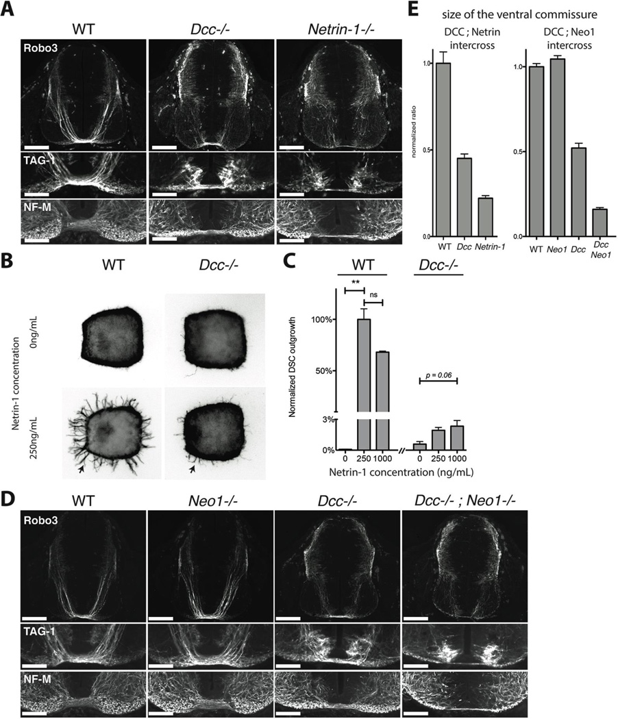
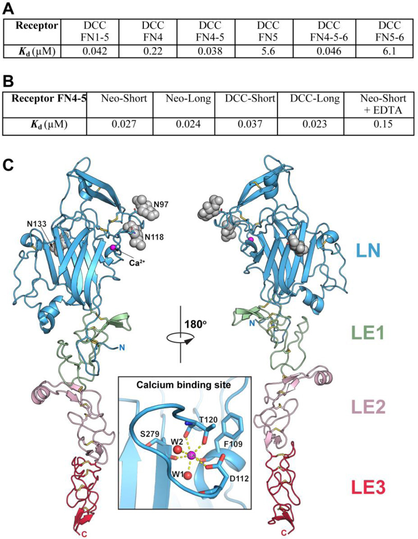
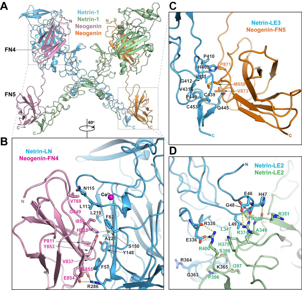
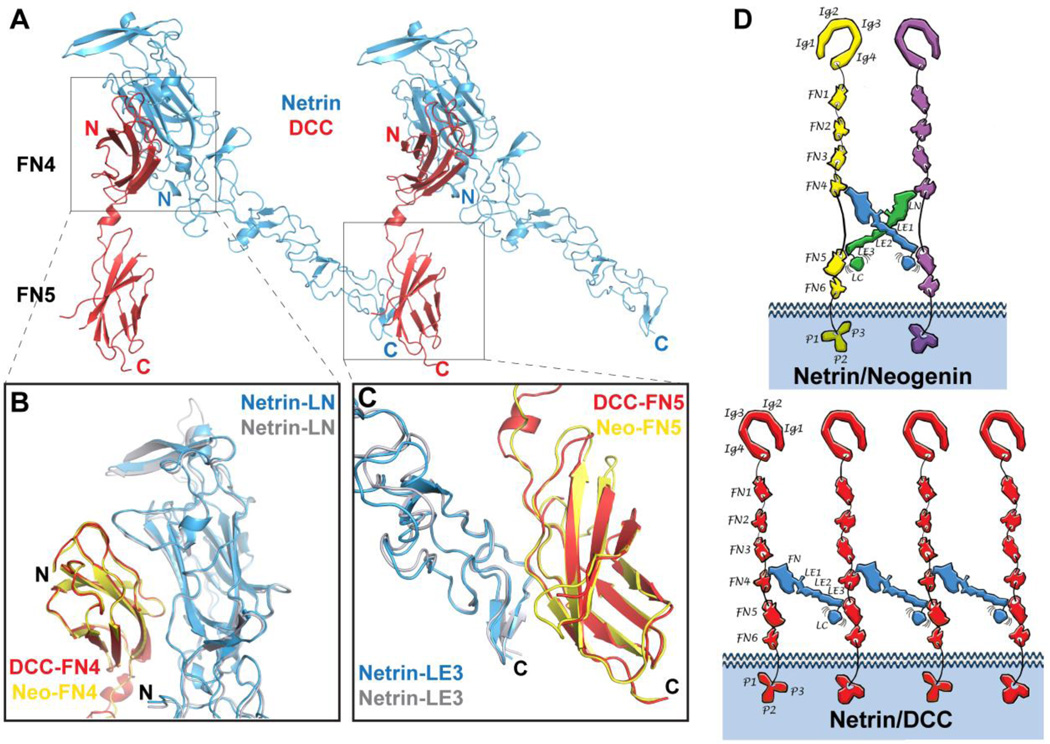
Netrins are secreted proteins that regulate axon guidance and neuronal migration. Deleted in colorectal cancer (DCC) is a well-established netrin-1 receptor mediating attractive responses. We provide evidence that its close relative neogenin is also a functional netrin-1 receptor that acts with DCC to mediate guidance in vivo. We determined the structures of a functional netrin-1 region, alone and in complexes with neogenin or DCC. Netrin-1 has a rigid elongated structure containing two receptor-binding sites at opposite ends through which it brings together receptor molecules. The ligand/receptor complexes reveal two distinct architectures: a 2:2 heterotetramer and a continuous ligand/receptor assembly. The differences result from different lengths of the linker connecting receptor domains fibronectin type III domain 4 (FN4) and FN5, which differs among DCC and neogenin splice variants, providing a basis for diverse signaling outcomes.


Figures

Fig. 1
Fig. 1
A) Cross sections of E11 wild-type, Dcc−/− and Netrin-1−/− littermate mouse embryos at the level of brachial spinal ganglia, stained for TAG-1, Robo3 and Neurofilament, Medium Chain (NF-M). Lower panels show details of the ventral commissural axon bundle. Dcc mutants have a reduced ventral commissure, but a large number of axons still cross. Netrin-1−/− embryos have a much-reduced ventral commissure. B) Axon outgrowth (arrows) in E11 wild type or Dcc−/− littermate mouse dorsal spinal cord explants cultured in 3D collagen gels with increasing concentrations of Netrin-1 and stained for Tuj1. C) Quantification of the response of axon outgrowth from wild type and Dcc−/− dorsal spinal cord explants to increasing Netrin-1 concentrations, normalized to wild type at 250ng/mL of Netrin-1. Dcc−/− mutants show a residual response to Netrin-1 application (arrow). D) Cross sections of E11 wild-type, Dcc−/−, Neo1−/− and Dcc−/−; Neo1−/− littermate mouse embryos at the level of brachial spinal ganglia, stained for TAG-1, Robo3 and Neurofilament (NF-M). Lower panels show details of the motor column and the ventral commissural axon bundle. The Dcc−/−; Neo1−/− double mutant has a much reduced ventral commissure and numerous axons in the motor column. E) Ratio of the commissural axon bundle size to the dorso-ventral spinal cord length of wild-type, Dcc−/− and Netrin1−/− embryos, normalized to wild types (left graph). Ratio of commissural axon bundle size to the dorso-ventral spinal cord length of Dcc−/−, Neo1−/− and Dcc−/−; Neo1−/− embryos normalized to wild type (right graph).The quantification shows the mean and SEM of 5 sections taken in brachial spinal cord in littermates, and is representative of 3 litters. Scale bars are 200µm (Robo3) and 100µm (TAG-1 and NF-M).
Fig. 2
Fig. 2
A) Binding of Netrin-1 (LN-LE1-LE2-LE3) to different DCC constructs documenting that the receptor FN4–FN5 region is necessary and sufficient for netrin binding. Kd, dissociation constant (in µM). B) Binding of Netrin-1 (LN-LE1-LE2-LE3) to the FN4–FN5 region of the different neogenin and DCC isoforms (see Fig. S4). To evaluate the role of the netrin-bound Ca++, 10 mM EDTA was added in one of the measurements. C) Structure of unbound Netrin-1. The individual netrin domains are labeled and colored in blue (LN), green (LE1), pink (LE2) and red (LE3). The glycosylation moieties at the three glycosylated Asn residues are shown as grey spheres. The N- and C- termini are labeled. The insert is a close-up view of the calcium-binding sire in the LN domain. The calcium ion is drawn in magenta and two bound water molecules – in red.
Fig. 3
Fig. 3
Structure of the Netrin-1/neogenin complex. A) Structure of the 2:2 Netrin-1/neogenin complex. The netrin molecules are in green and blue and the neogenin – in orange and magenta. The N- and C- termini of the molecules are labeled. B) Close-up view of the netrin-LN/neogenin-FN4 interface (Interface-1). Interacting residues and the netrin-bound calcium are labeled. C) Close-up view of the netrin-LE3/neogenin-FN5 interface (Interface-2). Interacting residues are labeled. D) Close-up view of the netrin-LE2/ netrin-LE2 interface (Interface-3). Interacting residues are labeled.
Fig. 4
Fig. 4
Structure of the Netrin-1/DCC complex. A) Structure of the Netrin-1/DCC complex. Netrin-1 is colored in blue, and neogenin in yellow and magenta. B) Superimposition of the LN-FN4 interaction site (Interface-1) in the Netrin-1/DCC (blue/yellow) and Netrin-1/neogenin (grey/pink) complexes. C) Superimposition of the LE3-FN5 interaction site (Interface-2) in the Netrin-1/DCC (blue/magenta) and Netrin-1/neogenin (grey/orange) complexes. D) Schematic drawing comparing the distinct Netrin-1/neogenin and Netrin-1/DCC signaling assemblies. The individual netrin and receptor domains are labeled. Ig, immunoglobulin; FN, fibronectin; LN, laminin-like N-terminal; LE, laminin-like EGF; LC, laminin-like C-terminal. Top, schematic representation if the 2:2 Netrin-1/neogenin complex. The netrin molecules are colored in blue and green, and the neogenin – in yellow and magenta. Bottom, schematic representation if the continuous Netrin-1/DCC assembly. The netrin molecules are colored in blue, and the DCC – in red. Interestingly, in both Netrin-1/DCC and Netrin-1/neogenin assemblies, the positively-charged netrin LC domain would be positioned towards the negatively-charged plasma membrane, thus potentially further stabilizing the signaling complexes at the neuronal surface.

Similar articles

Cited by

References

    1. Serafini T, Kennedy TE, Galko MJ, Mirzayan C, Jessell TM, Tessier-Lavigne M. The netrins define a family of axon outgrowth-promoting proteins homologous to C. elegans UNC-6. Cell. 1994;78:409–424. - PubMed
    1. Serafini T, Colamarino SA, Leonardo ED, Wang H, Beddington R, Skarnes WC, Tessier-Lavigne M. Netrin-1 is required for commissural axon guidance in the developing vertebrate nervous system. Cell. 1996;87:1001–1014. - PubMed
    1. Forcet C, Stein E, Pays L, Corset V, Llambi F, Tessier-Lavigne M, Mehlen P. Netrin-1-mediated axon outgrowth requires deleted in colorectal cancer-dependent MAPK activation. Nature. 2002;417:443–447. - PubMed
    1. Moore SW, Tessier-Lavigne M, Kennedy TE. Netrins and their receptors. Advances in experimental medicine and biology. 2007;621:17–31. - PubMed
    1. Rajasekharan S, Kennedy TE. The netrin protein family. Genome biology. 2009;10:239. - PMC - PubMed

Publication types

MeSH terms