Review
. 2014 Oct;34(10):2191-8.
doi: 10.1161/ATVBAHA.114.303422. Epub 2014 May 29.

Shear stress-initiated signaling and its regulation of endothelial function

Affiliations
Review

Shear stress-initiated signaling and its regulation of endothelial function

Jing Zhou et al. Arterioscler Thromb Vasc Biol. 2014 Oct.

Abstract

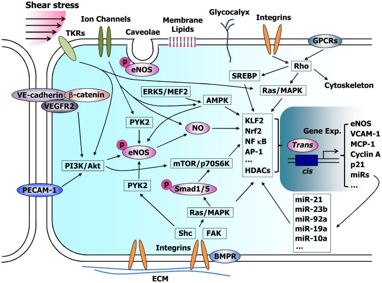
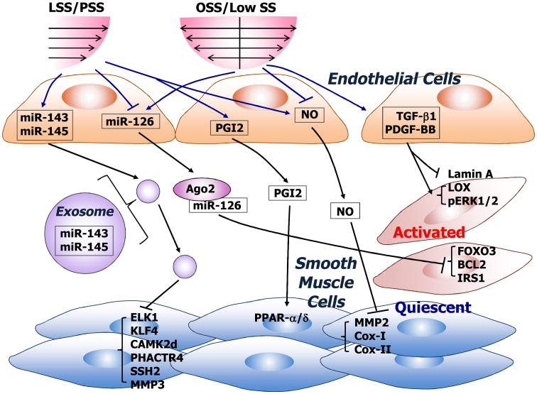
Atherosclerosis develops preferentially at branches and curvatures of the arterial tree, where blood flow pattern is disturbed rather than being laminar, and wall shear stress has an irregular distribution without defined directions. The endothelium in the atherosusceptible regions, in comparison to that in atheroresistant regions, shows activation of proproliferative and proinflammatory gene expressions, reduced production of nitric oxide (NO), increased leukocyte adhesion, and permeability, as well as other atheroprone phenotypes. Differences in gene expressions and cell phenotypes have been detected in endothelia residing in native atherosusceptible and atheroresistant regions of the arteries, or in arteries from animal models with artificial creation of disturbed flow. Similar results have also been shown in in vitro systems that apply controlled shear stresses with or without clear directions to cultured endothelial cells in fluid-dynamically designed flow-loading devices. The available evidence indicates that the coordination of multiple signaling networks, rather than individual separate pathways, links the mechanical signals to specific genetic circuitries in orchestrating the mechanoresponsive networks to evoke comprehensive genetic and functional responses.

Keywords: atherosclerosis; cellular mechanotransduction; hemodynamics.


Figures

Figure 1
Figure 1
Schematic diagram showing endothelial mechanotransduction and signaling induced by shear stress. Shear stress stimulates endothelial cells (ECs) through the activation of mechanosensors, including integrins, tyrosine kinase receptors (TKRs), G proteins and G protein-coupled receptors (GPCRs), ion channels, intercellular junction proteins (e.g., VE-cadherin and PECAM-1), caveolae, membrane lipids, and glycocalyx. These mechanosensors act through adaptor molecules (e.g., Shc) to trigger the activation of signaling molecules such as Ras, Rho, phosphatidylinositol-3-kinase (PI3K), and mitogen-activated protein kinases (MAPKs), which then activate eNOS, Smad1/5, and the transcription factors and cofactors (e.g., KLF2, NF kB, and AP-1) to regulate the expression of a number of functional genes such as eNOS, VCAM-1, and MCP-1, as well as microRNAs (miRs). This diagram illustrates that multiple signaling pathways coordinate to form mechanoresponsive networks to modulate EC phenotype and function.
Figure 2
Figure 2
Schematic diagram showing the modulation of gene expression and phenotype of smooth muscle cells (SMCs) by shear stress via ECs. Shear stress with sufficient magnitude and clear directions, i.e., laminar shear stress (LSS) and pulsatile shear stress (PSS), differ from that with a low magnitude and no clear direction, i.e., low shear stress and oscillatory shear stress (OSS) in their regulation of the release of secretory molecules such as NO, prostacyclin (PGI2), PDGF-BB and TGF-b1, and miRs. The uptake of NO, PGI2, and exosome-embedded miR-143/-145 by SMCs leads to functional targeting in SMCs with the promotion of a quiescent phenotype. In contrast, the PDGF-BB, TGF-b1, and Argonaute 2 (Ago2)-carried miR-126 released by ECs subjected to low shear stress or OSS induce an activated phenotype of SMCs.

Similar articles

Cited by

References

    1. Chien S. Mechanotransduction and endothelial cell homeostasis: the wisdom of the cell. Am J Physiol Heart Circ Physiol. 2007;292(3):H1209–1224. - PubMed
    1. Davies PF, Civelek M, Fang Y, Fleming I. The atherosusceptible endothelium: endothelial phenotypes in complex haemodynamic shear stress regions in vivo. Cardiovasc Res. 2013;99(2):315–327. - PMC - PubMed
    1. Davies PF. Hemodynamic shear stress and the endothelium in cardiovascular pathophysiology. Nat Clin Pract Cardiovasc Med. 2009;6(1):16–26. - PMC - PubMed
    1. Chiu JJ, Chien S. Effects of disturbed flow on vascular endothelium: pathophysiological basis and clinical perspectives. Physiol Rev. 2011;91(1):327–387. - PMC - PubMed
    1. Won D, Zhu SN, Chen M, Teichert AM, Fish JE, Matouk CC, Bonert M, Ojha M, Marsden PA, Cybulsky MI. Relative reduction of endothelial nitric-oxide synthase expression and transcription in atherosclerosis-prone regions of the mouse aorta and in an in vitro model of disturbed flow. Am J Pathol. 2007;171(5):1691–1704. - PMC - PubMed

Publication types