. 2014 Jul 25;289(30):20858-70.
doi: 10.1074/jbc.M114.566687.

Mammalian COPII coat component SEC24C is required for embryonic development in mice

Mammalian COPII coat component SEC24C is required for embryonic development in mice

Elizabeth J Adams et al. J Biol Chem. .

Abstract

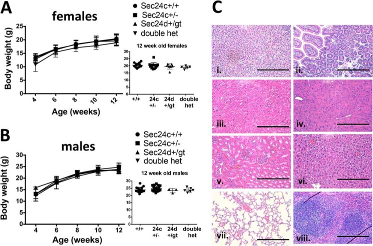
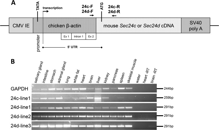
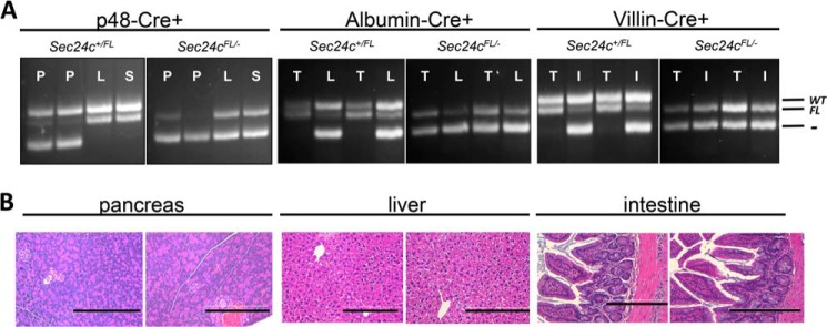
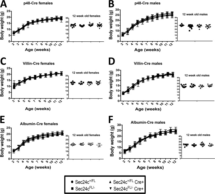
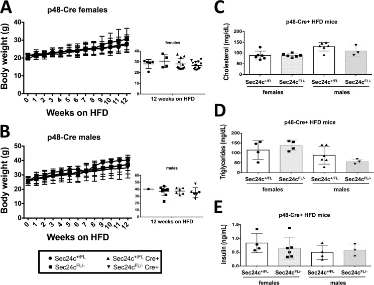
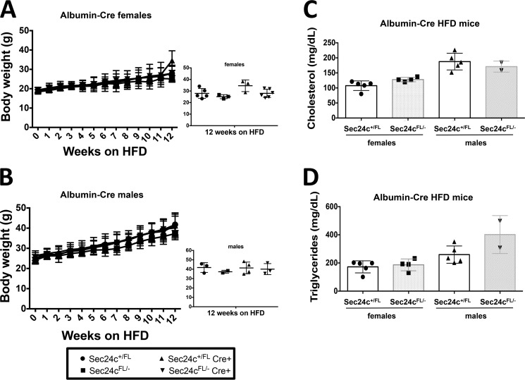
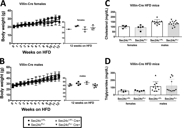
COPII-coated vesicles mediate the transport of newly synthesized proteins from the endoplasmic reticulum to the Golgi. SEC24 is the COPII component primarily responsible for recruitment of protein cargoes into nascent vesicles. There are four Sec24 paralogs in mammals, with mice deficient in SEC24A, -B, and -D exhibiting a wide range of phenotypes. We now report the characterization of mice with deficiency in the fourth Sec24 paralog, SEC24C. Although mice haploinsufficient for Sec24c exhibit no apparent abnormalities, homozygous deficiency results in embryonic lethality at approximately embryonic day 7. Tissue-specific deletion of Sec24c in hepatocytes, pancreatic cells, smooth muscle cells, and intestinal epithelial cells results in phenotypically normal mice. Thus, SEC24C is required in early mammalian development but is dispensable in a number of tissues, likely as a result of compensation by other Sec24 paralogs. The embryonic lethality resulting from loss of SEC24C occurs considerably later than the lethality previously observed in SEC24D deficiency; it is clearly distinct from the restricted neural tube phenotype of Sec24b null embryos and the mild hypocholesterolemic phenotype of adult Sec24a null mice. Taken together, these results demonstrate that the four Sec24 paralogs have developed unique functions over the course of vertebrate evolution.


Figures

FIGURE 1.
FIGURE 1.
Identification of an alternative splice form of Sec24c. A, schematic of the two Sec24c splice forms observed in mice. Arrows indicate primers used to detect the presence of the alternative exon (6*). Shaded regions in exons 4 and 5 represent exonic sequence present in Sec24c-2 that is absent from another RefSeq splice variant annotated as containing exon 6* (NM_001168273.1), which was not detected in our RT-PCR. B, protein sequence alignment beginning at Thr-306 of SEC24C-1 and SEC24C-2 showing the additional 23 amino acids encoded by exon 6*. C, RT-PCR using primers V and W, which detects a 249-bp product from Sec24c-2 and a 180-bp product from Sec24c-1.
FIGURE 2.
FIGURE 2.
Generation of Sec24c conditional and null alleles. A, schematic of the original Sec24cGT allele. FLPe-mediated excision yields the conditional Sec24cFL allele, and subsequent Cre-mediated excision gives rise to the null Sec24c allele. Primers used for genotyping are indicated. B, long range PCR confirms correct targeting for the original Sec24cGT allele. Primers GF3 and GR4 are located outside of the homology arms. Primers GF3 and B amplify a 6234-bp product from the 5′ end of the Sec24cGT allele, and primers G and GR4 amplify a 6648-bp product from the 3′ end. As expected, neither set of primers yields a product from the Sec24cWT allele. C, genotyping with primers A, Ex, and E distinguishes between the wild type (308 bp), gene trap or floxed (278 bp), and null alleles (225 bp).
FIGURE 3.
FIGURE 3.
Phase contrast (A–C) and H&E staining (D–F) of E7.5 embryos resulting from Sec24c+/− intercrosses. E7.5 embryos were removed from the decidua and photographed prior to fixation (A–C), or whole decidual swellings were fixed and sectioned (D–F). Images are representative of the types of embryonic development/loss observed in 22 embryos. A, B, and D illustrate normal gastrulation. Genotypes were obtained for A–C (A, Sec24c+/+; B, Sec24c+/−; C, Sec24c−/−). The embryo in C exhibits thinning of the embryonic ectoderm. E illustrates a disorganized embryo, and F shows remnants of the egg cylinder. Anterior is to the left of each embryo; A = amniotic fold; EPC = ectoplacental cone; PS = primitive streak. Arrow in F indicates remnants of the embryo.
FIGURE 4.
FIGURE 4.
Sec24c+/− mice are phenotypically normal. Growth curves of female (A) and male (B) mice indicate no difference in body weights between heterozygous mice and wild type littermate controls, with p > 0.05 at all time points. Error bars represent standard deviation. C, H&E staining of Sec24c+/− tissues reveals no abnormalities in a panel of tissues, including adrenal gland (panel i), intestine (panel ii), heart (panel iii), pancreas (panel iv), kidney (panel v), liver (panel vi), lung (panel vii), and spleen (panel viii). Scale bar, 0.25 μm.
FIGURE 5.
FIGURE 5.
Sec24c+/−Sec24d+/GT mice are phenotypically normal. Growth curves of female (A) and male (B) mice indicate no difference in body weights between heterozygous mice and littermate controls, with p > 0.05 at all time points. Error bars represent standard deviation. “Double het” = Sec24c+/− Sec24d+/GT. C, H&E staining of Sec24c+/− Sec24d+/GT tissues does not reveal any abnormalities in a panel of tissues, including adrenal gland (panel i), intestine (panel ii), heart (panel iii), pancreas (panel iv), kidney (panel v), liver (panel vi), lung (panel vii), and spleen (panel viii). Scale bar, 0.25 μm.
FIGURE 6.
FIGURE 6.
Ubiquitous transgenic expression of Sec24c and Sec24d. A, diagram of the pCAG3zS-Sec24c or -Sec24d transgenic construct, adapted from Ref. , including transcription start site, initial ATG, and polyadenylation signal. CMV IE = cytomegalovirus immediate-early enhancer; UTR = untranslated region; SV40 poly(A) = simian virus 40 polyadenylation signal. Primers 24c-F, 24c-R, 24d-F, and 24d-R were used for RT-PCR of transgenes expressing Sec24c or Sec24d, respectively. B, transgene expression in a panel of tissues for founder lines 24c-line1, 24d-line1, 24d-line2, and 24d-line3. Heart and brain RT samples were carried out in the absence of reverse transcriptase.
FIGURE 7.
FIGURE 7.
Tissue-specific Cre-mediated excision of Sec24cFL allele. A, genotyping for Cre-mediated excision in the pancreas (P), liver (L), spleen (S), intestine (I), and tail (T) in Sec24c+/FL and Sec24cFL/− mice carrying the p48-Cre, albumin-Cre, or villin-Cre transgenes. The floxed allele is excised to generate the null allele in the pancreas, liver, or intestine, respectively, but is preserved in other tissues. The sizes of the expected products for wild type (WT), flox (FL), and null (−) alleles are indicated. B, H&E staining of pancreatic tissue from p48-Cre+ mice, liver tissue from albumin-Cre+ mice, and intestine from villin-Cre+ mice show no abnormalities. Scale bar, 0.25 μm.
FIGURE 8.
FIGURE 8.
Loss of SEC24C in the pancreas, liver, or intestine has no effect on growth. Body weight analysis over 12 weeks of Sec24cFL/−p48-Cre+ mice (A and B), Sec24cFL/− albumin-Cre+ mice (C and D), and Sec24cFL/−villin-Cre+ mice (E and F) with corresponding littermate controls in females and males. Insets show individual body weights at 12 weeks. p > 0.05 at all time points. Error bars represent standard deviation.
FIGURE 9.
FIGURE 9.
Sec24cFL/−p48-Cre+ mice display expected diet-induced obesity. Sec24cFL/−p48-Cre+ and littermate controls show no difference in weight gain over 12 weeks on HFD for females (A) and males (B), with p > 0.05 at all time points. Cholesterol levels (C), triglyceride levels (D), and insulin levels (E) are indistinguishable from littermate controls after 12 weeks on HFD. All p values >0.05 and error bars represent standard deviation.
FIGURE 10.
FIGURE 10.
Sec24cFL/−albumin-Cre+ mice display expected diet-induced obesity. Sec24cFL/−albumin-Cre+ and littermate controls show no difference in weight gain over 12 weeks on HFD for females (A) and males (B), with p > 0.05 at all time points. Cholesterol levels (C) and triglyceride levels (D) are indistinguishable from littermate controls after 12 weeks on HFD. All p values >0.05 and error bars represent standard deviation.
FIGURE 11.
FIGURE 11.
Sec24cFL/−villin-Cre+ mice display expected diet-induced obesity. Sec24cFL/−villin-Cre+ and littermate controls show no difference in weight gain over 12 weeks on HFD for females (A) and males (B), with p > 0.05 at all time points. Cholesterol levels (C) and triglyceride levels (D) are indistinguishable from littermate controls after 12 weeks on HFD. All p values >0.05 and error bars represent standard deviation.

References

    1. Bonifacino J. S., Glick B. S. (2004) The mechanisms of vesicle budding and fusion. Cell 116, 153–166 - PubMed
    1. Palade G. (1975) Intracellular aspects of the process of protein synthesis. Science 189, 867–867 - PubMed
    1. Budnik A., Stephens D. J. (2009) ER exit sites–localization and control of COPII vesicle formation. FEBS Lett. 583, 3796–3803 - PubMed
    1. Lee M. C., Miller E. A. (2007) Molecular mechanisms of COPII vesicle formation. Semin. Cell Dev. Biol. 18, 424–434 - PubMed
    1. Zanetti G., Pahuja K. B., Studer S., Shim S., Schekman R. (2012) COPII and the regulation of protein sorting in mammals. Nat. Cell Biol. 14, 20–28 - PubMed

Publication types

Substances