

Age- and species-dependent infiltration of macrophages into the testis of rats and mice exposed to mono-(2-Ethylhexyl) phthalate (MEHP)
- PMID: 24876407
- PMCID: PMC4434960
- DOI: 10.1095/biolreprod.113.115527
Age- and species-dependent infiltration of macrophages into the testis of rats and mice exposed to mono-(2-Ethylhexyl) phthalate (MEHP)
Abstract
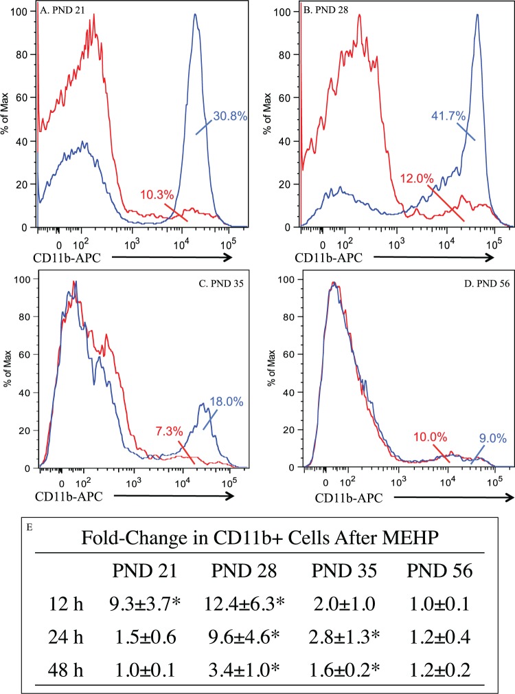
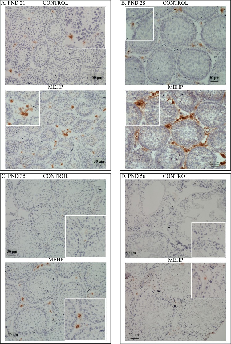
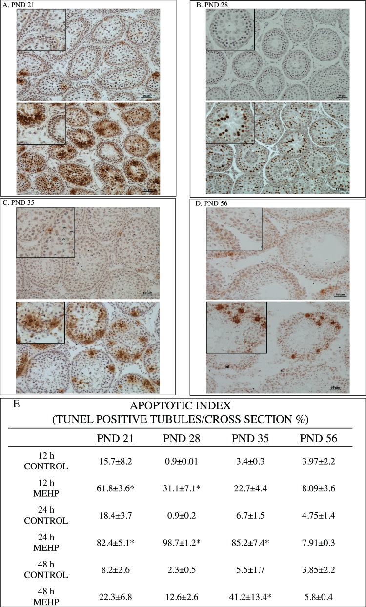
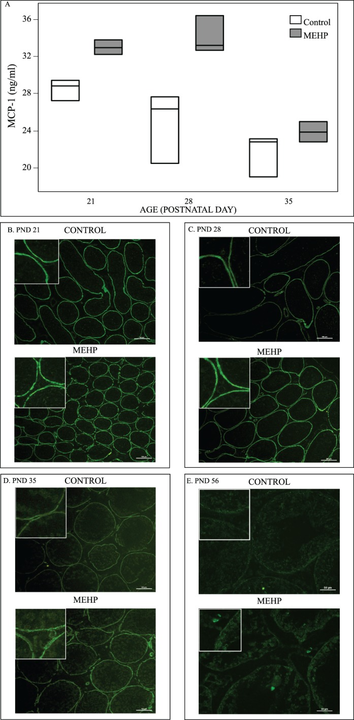
The mechanism by which noninfectious testicular inflammation results in infertility is poorly understood. Here the infiltration of CD11b+ immunoreactive testicular interstitial cells (neutrophil, macrophages, dendritic cells) in immature (Postnatal Day [PND] 21, 28, and 35) and adult (PND 56) Fischer rats is described at 12, 24, and 48 h after an oral dose of 1 g/kg mono-(2-ethylhexyl) phthalate (MEHP), a well-described Sertoli cell toxicant. Increases of CD11b+ cells are evident 12 h after MEHP exposure in PND 21 and 28 rats. In PND 28 rats, CD11b+ cells remained significantly elevated at 48 h, while in PND 21 rats, it returned to control levels by 24 h. The peak number of CD11b+ cells in PND 35 rat testis is delayed until 24 h, but remains significantly elevated at 48 h. In PND 56 rats, no increase in CD11b+ cells occurs after MEHP exposure. In PND 21, 28, and 35 rats, a significant increase in monocyte chemoattractant protein-1 (MCP-1) by peritubular myoid cells occurs 12 h after MEHP. Interestingly, MEHP treatment of C57BL/6J mice did not incite an infiltration of CD11b+ cells at either PND 21 or 28. The peak level of germ cell apoptosis observed 24 h after MEHP exposure in young rats is not seen in mice at any age or in PND 56 rats. Taken together, these findings implicate MCP-1 released by peritubular myoid cells in provoking the migration of CD11b+ cells into the immature rat testis early after MEHP exposure and point to a role for CD11b+ cells in triggering germ cell apoptosis in an age- and species-dependent manner.
Keywords: interstitial cells; macrophage; myoid cells; reproductive immunology; toxicology.
© 2014 by the Society for the Study of Reproduction, Inc.
Figures


Similar articles
-
Mono-(2-ethylhexyl) phthalate-induced Sertoli cell injury stimulates the production of pro-inflammatory cytokines in Fischer 344 rats.Reprod Toxicol. 2017 Apr;69:150-158. doi: 10.1016/j.reprotox.2017.02.013. Epub 2017 Feb 24. Reprod Toxicol. 2017. PMID: 28238932 Free PMC article.
-
Peritubular myoid cells of the testis produce monocyte chemotactic protein 1 upon direct exposure to Mono-(2-Ethylhexyl) phthalate through the IL-1 signaling pathway.Toxicology. 2025 Jun;514:154118. doi: 10.1016/j.tox.2025.154118. Epub 2025 Mar 13. Toxicology. 2025. PMID: 40089264
-
MEHP-induced rat testicular inflammation does not exacerbate germ cell apoptosis.Reproduction. 2018 Jul;156(1):35-46. doi: 10.1530/REP-18-0093. Epub 2018 May 9. Reproduction. 2018. PMID: 29743262 Free PMC article.
-
FasL gene-deficient mice display a limited disruption in spermatogenesis and inhibition of mono-(2-ethylhexyl) phthalate-induced germ cell apoptosis.Toxicol Sci. 2010 Apr;114(2):335-45. doi: 10.1093/toxsci/kfq015. Epub 2010 Jan 25. Toxicol Sci. 2010. PMID: 20100735 Free PMC article.
-
Responses of peritubular macrophages and the testis transcriptome profiles of peripubertal and adult rodents exposed to an acute dose of MEHP.Toxicol Sci. 2024 Feb 28;198(1):76-85. doi: 10.1093/toxsci/kfad128. Toxicol Sci. 2024. PMID: 38113427 Free PMC article.
Cited by
-
Peritubular Macrophages Are Recruited to the Testis of Peripubertal Rats After Mono-(2-Ethylhexyl) Phthalate Exposure and Is Associated With Increases in the Numbers of Spermatogonia.Toxicol Sci. 2021 Aug 3;182(2):288-296. doi: 10.1093/toxsci/kfab059. Toxicol Sci. 2021. PMID: 34010400 Free PMC article.
-
The Impact of Chronic Phthalate Exposure on Rodent Anxiety and Cognition.Biol Psychiatry Glob Open Sci. 2023 Jul 21;4(1):203-212. doi: 10.1016/j.bpsgos.2023.07.002. eCollection 2024 Jan. Biol Psychiatry Glob Open Sci. 2023. PMID: 38298799 Free PMC article. Review.
-
Mono-(2-ethylhexyl) phthalate-induced Sertoli cell injury stimulates the production of pro-inflammatory cytokines in Fischer 344 rats.Reprod Toxicol. 2017 Apr;69:150-158. doi: 10.1016/j.reprotox.2017.02.013. Epub 2017 Feb 24. Reprod Toxicol. 2017. PMID: 28238932 Free PMC article.
-
Effect of prenatal DINCH plasticizer exposure on rat offspring testicular function and metabolism.Sci Rep. 2017 Sep 11;7(1):11072. doi: 10.1038/s41598-017-11325-7. Sci Rep. 2017. PMID: 28894178 Free PMC article.
-
Regulation of microtubule (MT)-based cytoskeleton in the seminiferous epithelium during spermatogenesis.Semin Cell Dev Biol. 2016 Nov;59:35-45. doi: 10.1016/j.semcdb.2016.01.004. Epub 2016 Jan 11. Semin Cell Dev Biol. 2016. PMID: 26791048 Free PMC article. Review.
References
-
- Albro PW, Jordan S, Corbett JT, Schroeder JL. Determination of total phthalate in urine by gas chromatography. Anal Chem. 1984;56:247–250. - PubMed
-
- Sjoberg P, Lindquist NG, Montin G, Ploen L. Effects of repeated intravenous infusions of the plasticizer di-(2-ethylhexyl) phthalate in young male rats. Arch Toxicol. 1985;58:78–83. - PubMed
-
- Tomita I, Nakamura Y, Yagi Y. Phthalic acid esters in various foodstuffs and biological materials. Ecotoxicol Environ Saf. 1977;1:275–287. - PubMed
-
- Boekelheide K, Johnson KJ, Richburg J. Sertoli cell toxicants In: Skinner MK, Griswold MD. (eds.), Sertoli Cell Biology. San Diego, CA: Elsevier Academic Press; 2005: 345 382
-
- Wittassek M, Koch HM, Angerer J, Bruning T. Assessing exposure to phthalates—the human biomonitoring approach. Mol Nutr Food Res. 2011;55:7–31. - PubMed
Publication types
MeSH terms
Substances
Grants and funding
LinkOut - more resources
Full Text Sources
Other Literature Sources
Research Materials
Miscellaneous
