

Bascule caecal volvulus: a rare cause of intestinal obstruction
- PMID: 24876459
- PMCID: PMC3998522
- DOI: 10.1093/jscr/rju025
Bascule caecal volvulus: a rare cause of intestinal obstruction
Abstract
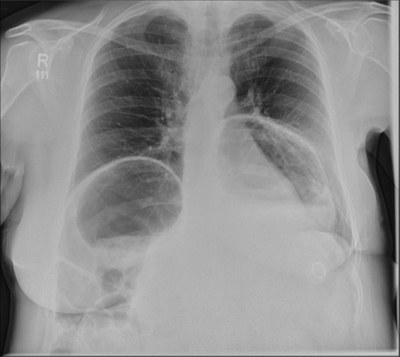
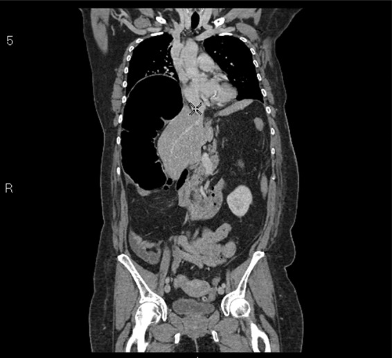
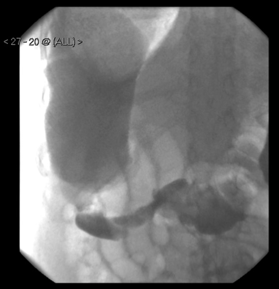
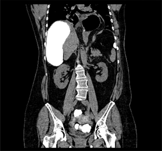
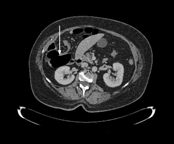
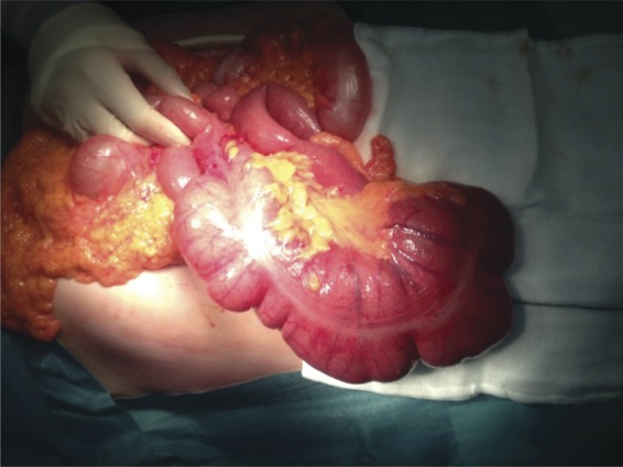
Caecal volvulus is a rare cause of intestinal obstruction, with the bascule subtype accounting for <10% of all cases of caecal volvulus. It is associated with significant morbidity and mortality if left undiagnosed. We present the case of a 58-year-old female who presented to our surgical department with symptoms of intestinal obstruction. She had various radiological investigations, which supported the diagnosis of a caecal volvulus of the bascule subtype. She was subsequently managed surgically and had a right hemicolectomy and ileocolic anastomosis. Her recovery was uneventful and she was discharged within 1 week of having her operation. Fortunately, caecal volvulus of the bascule subtype is rarely encountered; however, clinicians need to be aware of its presentation and subsequent management options so that clinical outcomes are improved.
Published by Oxford University Press and JSCR Publishing Ltd. All rights reserved. © The Author 2014.
Figures
Similar articles
-
Caecal bascule - A variant of caecal volvulus: Review of diagnostic challenges and approaches.Surgeon. 2022 Oct;20(5):e262-e265. doi: 10.1016/j.surge.2021.09.004. Epub 2021 Nov 14. Surgeon. 2022. PMID: 34789426 Review.
-
Caecal bascule: a case series and literature review.ANZ J Surg. 2018 May;88(5):E386-E389. doi: 10.1111/ans.13898. Epub 2017 Mar 20. ANZ J Surg. 2018. PMID: 28318090 Review.
-
[Caecal bascule, an unusual cause of intestinal obstruction].Cir Cir. 2016 Nov-Dec;84(6):513-517. doi: 10.1016/j.circir.2015.10.004. Epub 2015 Dec 11. Cir Cir. 2016. PMID: 26688476 Spanish.
-
Complete common mesentery revealed by a cecal bascule: Two case reports.Int J Surg Case Rep. 2025 Apr;129:111160. doi: 10.1016/j.ijscr.2025.111160. Epub 2025 Mar 16. Int J Surg Case Rep. 2025. PMID: 40106949 Free PMC article.
-
Caecal Volvulus, Untwisting the Twisted: A Case Report and Literature Review.Cureus. 2022 Nov 1;14(11):e30961. doi: 10.7759/cureus.30961. eCollection 2022 Nov. Cureus. 2022. PMID: 36465206 Free PMC article.
Cited by
-
An Unlikely Cause of Abdominal Pain.Clin Pract Cases Emerg Med. 2018 Mar 14;2(2):139-142. doi: 10.5811/cpcem.2018.2.37073. eCollection 2018 May. Clin Pract Cases Emerg Med. 2018. PMID: 29849249 Free PMC article.
-
Cecal bascule as a cause of postoperative nausea and abdominal pain.Radiol Case Rep. 2019 Mar 27;14(6):697-699. doi: 10.1016/j.radcr.2019.02.010. eCollection 2019 Jun. Radiol Case Rep. 2019. PMID: 30976371 Free PMC article.
-
Uncomplicated Amyand's hernia in a setting of abdominal wall insufficiency: a case report.J Med Case Rep. 2025 Jan 13;19(1):15. doi: 10.1186/s13256-024-05015-y. J Med Case Rep. 2025. PMID: 39806483 Free PMC article.
-
Cecal bascule in pregnancy: a case report and review of the literature.J Surg Case Rep. 2023 May 22;2023(5):rjad287. doi: 10.1093/jscr/rjad287. eCollection 2023 May. J Surg Case Rep. 2023. PMID: 37234082 Free PMC article.
-
Cecal bascule after spinal cord injury: A case series report.Int J Surg Case Rep. 2016;22:94-7. doi: 10.1016/j.ijscr.2016.03.040. Epub 2016 Apr 1. Int J Surg Case Rep. 2016. PMID: 27077698 Free PMC article.
References
-
- Habre J, Sautot-Vial N, Marcotte C, Benchimol D. Caecal volvulus. Am J Surg. 2008;196:e48–9. - PubMed
-
- Rabinovici R, Simansky DA, Kaplan O, Mavor E, Manny J. Cecal volvulus. Dis Colon Rectum. 1990;33:765–9. - PubMed
-
- Madiba TE, Thomson SR. The management of cecal volvulus. Dis Colon Rectum. 2002;45:264–7. - PubMed
-
- Tejler G, Jiborn H. Volvulus of the cecum. Report of 26 cases and review of the literature. Dis Colon Rectum. 1988;31:445–9. - PubMed
Publication types
LinkOut - more resources
Full Text Sources
Other Literature Sources
