. 2014 May 30;344(6187):992-7.
doi: 10.1126/science.1251915.

Crystal structure of a heterotetrameric NMDA receptor ion channel

Affiliations

Crystal structure of a heterotetrameric NMDA receptor ion channel

Erkan Karakas et al. Science. .

Abstract

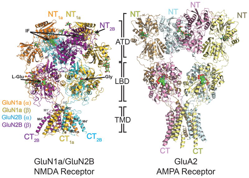
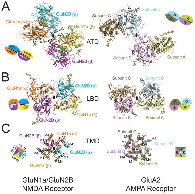
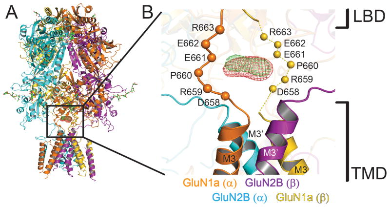
N-Methyl-D-aspartate (NMDA) receptors belong to the family of ionotropic glutamate receptors, which mediate most excitatory synaptic transmission in mammalian brains. Calcium permeation triggered by activation of NMDA receptors is the pivotal event for initiation of neuronal plasticity. Here, we show the crystal structure of the intact heterotetrameric GluN1-GluN2B NMDA receptor ion channel at 4 angstroms. The NMDA receptors are arranged as a dimer of GluN1-GluN2B heterodimers with the twofold symmetry axis running through the entire molecule composed of an amino terminal domain (ATD), a ligand-binding domain (LBD), and a transmembrane domain (TMD). The ATD and LBD are much more highly packed in the NMDA receptors than non-NMDA receptors, which may explain why ATD regulates ion channel activity in NMDA receptors but not in non-NMDA receptors.


Figures

Figure 1
Figure 1. Overall structure of heterotetrameric GluN1a/GluN2B NMDA receptor and comparison with GluA2 AMPA receptor
Overall structures of GluN1a/GluN2B NMDA receptor (left) and GluA2 AMPA receptor (right, PDB ID: 3KG2). Both structures are placed so that the tetramers of both receptors are in the similar orientation at the LBD layer. GluN1a and GluN2B subunits, labeled as GluN1a (α), GluN1a (β), GluN2B (α), GluN2B (β) are colored in orange, yellow, cyan and purple, respectively. The amino (NT) and carboxy (CT) termini are located on top and bottom, respectively. Ifenprodil (IF), located at the GluN1a/GluN2B ATD heterodimer interfaces, and agonists, glycine (Gly) and L-glutamate (L-Glu), lodged at the LBD clamshells, are shown in green spheres. N-glycosylation chains are shown in green sticks.
Figure 2
Figure 2. Domain-by-domain structural comparison of heteromeric GluN1a/GluN2B NMDA receptors and homomeric GluA2 AMPA receptor
Structures of ATD (A), LBD (B), and TMD (C) viewed from the top of the receptors. All of the domains are assembled around the overall two-fold axis (large black oval) in GluN1a/GluN2B heterotetramers (left). In GluA2 homotetramers (right), the local two-fold axis (small black oval) runs within the ATD and LBD dimers, a two-fold axis (large black oval) runs between the ATD and LBD dimers, and the fourfold axis (black square) runs in the center of the TMD. Schematic figures next to the structures represent subunit organization at each domain, where subunits with black dots in between represent dimer pairs. Ifenprodil, glycine, L-glutamate, and ZK200775 are shown in spheres. N-glycosylation chains are shown in green sticks.
Figure 3
Figure 3. Inter- and intra-subunit interfaces in GluN1a/GluN2B NMDA receptors
(A) Ribbon and surface representation of GluN1a/GluN2B NMDA receptor colored as in Fig. 1. Inter-subunit interfaces that are probed by disulfide cross-linking experiments are surrounded by colored boxes. (B) Western blot analysis of disulfide bond formation by cysteine substitutions at the subunit interfaces probed by anti-GluN1 (top) and anti-GluN2B (bottom) antibodies under non-reducing conditions. Arrows indicate positions of non-cross-linked monomers and cross-linked dimers and tetramers. (C–G) Close up views of the inter- and intra-subunit interfaces between GluN2B ATD (α) and GluN2B-ATD (β) (Site-I, yellow box) (C), between GluN1a ATD and GluN1a LBD, and GluN1a-ATD and GluN2B-LBD (Site-II, green box) (D), between GluN1a LBD and GluN2B ATD, and GluN2B ATD and GluN2B LBD (Site-III, blue box) (E), between GluN1a LBD and GluN2B LBD (Site-IV, red box) (F) and between GluN1a TMD and GluN2B TMD (Site-V, purple box) (G). Side chains without clear electron densities are modeled as alanine. Residues that are mutated to cysteine are labeled in red.
Figure 4
Figure 4. Structural comparison of NMDA receptor ion channel with GluA2 AMPA receptor and Shaker potassium channels
TMDs of GluN1a subunits (yellow, left panel) and GluN2B subunits (cyan, middle panel) are superposed onto the ion channel regions (red) of the closed conformation of GluA2 AMPA receptor (PDB ID: 3KG2) (A), open conformation of Shaker potassium channel (PDB ID: 2R9R) (B). The superposed structures are viewed from the side (left and middle panels) or from the extracellular side (right panel). Superposition is performed using Secondary-structure matching (SSM) tool in the program Coot. Loops are excluded from the figure for clarity.
Figure 5
Figure 5. Putative calcium binding site at the extracellular vestibule
(A) Overall structure of GluN1a/GluN2B NMDA receptors with the anomalous Fourier difference maps for holmium (green mesh; from the 7.5 Å dataset) and gadolinium (red mesh; from the 7.8 Å dataset) countered at 4.5 σ. (B) Close up view of the boxed region in panel A. Holmium and gadolinium binding sites are located at the extracellular vestibule over the bundle of M3 helices. Cα atoms of the residues on GluN1a DRPEER motif from the GluN1a/GluN2Bcrystx structure are shown as spheres. Residues for the disordered DRPEER motif (shown as dashed lines) on the GluN1a (β) protomer (yellow) are positioned based on the structural alignment of the GluN1a (α) protomer (orange).

References

    1. Hayashi T. Effects of sodium glutamate on the nervous system. Keio J Med. 1954;3:192.
    1. Mayer ML, Westbrook GL, Guthrie PB. Voltage-dependent block by Mg2+ of NMDA responses in spinal cord neurones. Nature. 1984 May 17–23;309:261. - PubMed
    1. Traynelis SF, et al. Glutamate receptor ion channels: structure, regulation, and function. Pharmacol Rev. 2010 Sep;62:405. - PMC - PubMed
    1. Gielen M, Siegler Retchless B, Mony L, Johnson JW, Paoletti P. Mechanism of differential control of NMDA receptor activity by NR2 subunits. Nature. 2009 Jun 4;459:703. - PMC - PubMed
    1. Yuan H, Hansen KB, Vance KM, Ogden KK, Traynelis SF. Control of NMDA receptor function by the NR2 subunit amino-terminal domain. The Journal of neuroscience. 2009 Sep 30;29:12045. - PMC - PubMed

Publication types

Associated data