

The Clinical, Radiographic and Histological evaluation of three different concentrations of Formocresol as a pulpotomy agent
- PMID: 24876712
- PMCID: PMC4037803
The Clinical, Radiographic and Histological evaluation of three different concentrations of Formocresol as a pulpotomy agent
Abstract
Background: Formocresol, though the center of much controversy is still the most widely used medicament for primary teeth pulpotomy and an intracanal medicament which has undergone a lengthy evolution to shorten the formocresol application time and reduce the concentration of formocresol exposure to the pulp tissue. Hence, the determination of the actual effective dose and concentration of formocresol for clinical application in primary teeth is an important area of research and a thorough clinical, radiographic and histological investigation in human subjects is very much needed.
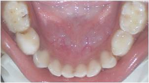
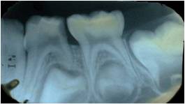
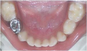
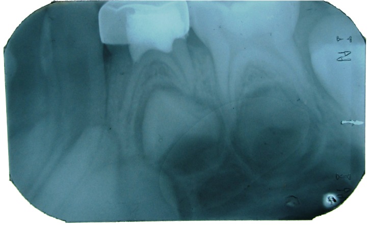
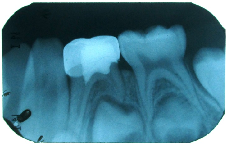
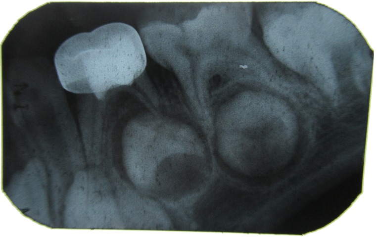
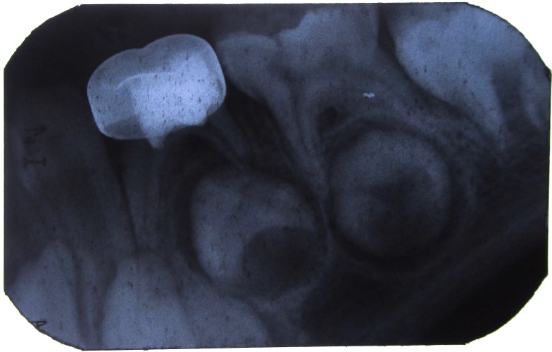
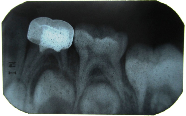
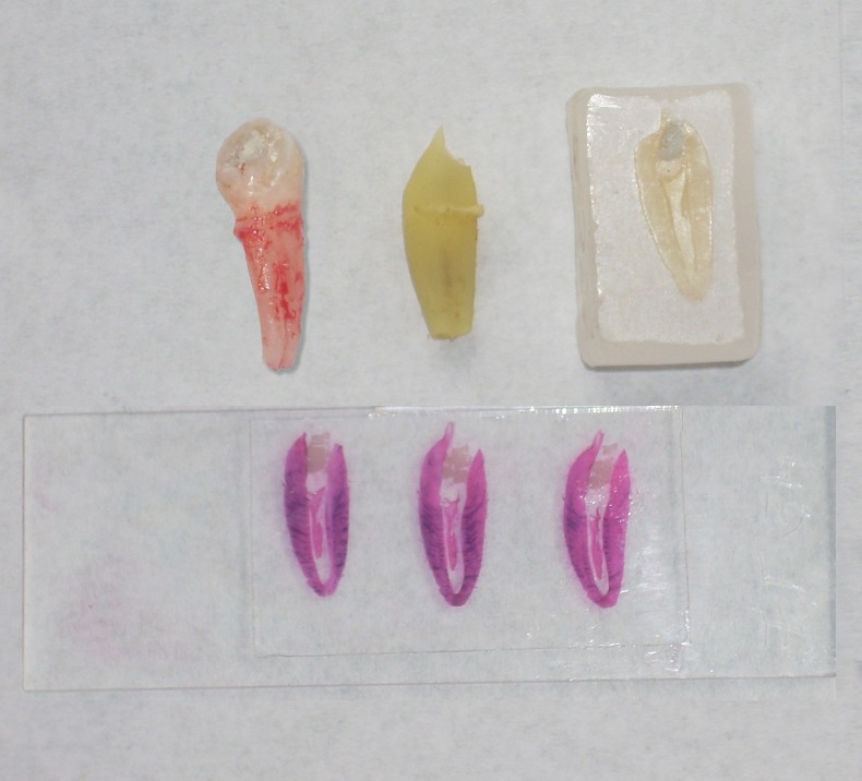
Materials & methods: The study was conducted on 45 primary molars for the Clinical, Radiographic study and 45 premolars orthodontically indicated for extraction for the Histological study. The samples were randomly and equally divided into 3 groups of 15 each for pulpotomy with full strength formocresol, 1:5 diluted formocresol and 1:25 diluted formocresol respectively. The pulpotomized primary molars were clinically evaluated at 1st, 3rd, 6th and 9th month while the pulpotomized premolars were subjected for histological evaluation after extraction.
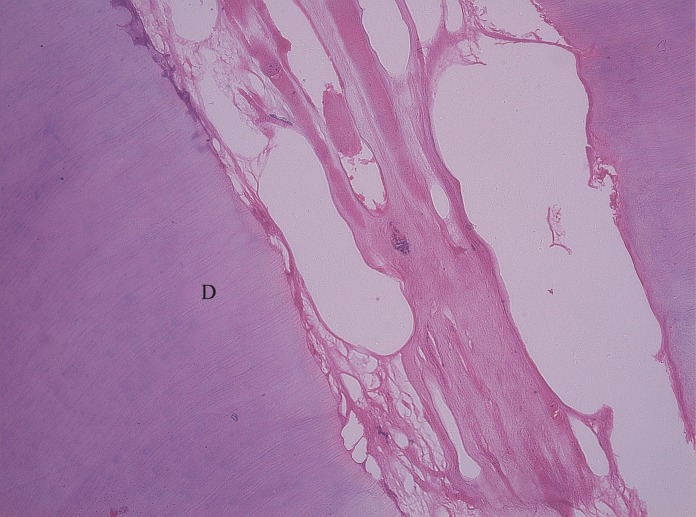
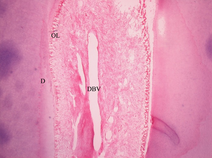
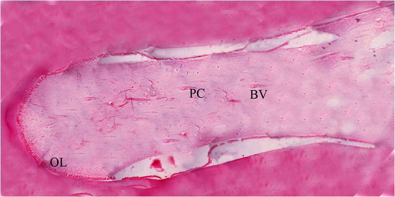
Results: Obtained by chi-square test revealed that all the pulpotomized primary molars were asymptomatic till the end of the study period; suggesting 100% clinical and radiographic success while histologically, the three concentrations of formocresol showed decreased severity of fixation of the pulp tissue with decreasing concentration of formocresol.
Conclusion: It can be inferred that the diluted formulations (1:5 and 1:25) of formocresol are equally efficient when compared to full-strength formocresol and thus, can be recommended for pulpotomy in primary teeth. How to cite the article: Goyal S, Abuwala T, Joshi K, Mehta J, Indushekar KR, Hallikerimath S. The Clinical, Radiographic and Histological evaluation of three different concentrations of Formocresol as a pulpotomy agent. J Int Oral Health 2014;6(2):118-25.
Keywords: Dilute formocresol; formocresol; primary teeth; pulpotomy.
Conflict of interest statement
Conflict of Interest: None
Figures

References
-
- JP Buckley. The chemistry of pulp decomposition, with a rational treatment for this condition & its sequelae. Am Dent J. 1904;3:764.
-
- D Zurn, NS Seale. Light-cured Calcium hydroxide vs Formocresol in Human Primary Molar Pulpotomies: A Randomized Controlled trial. Pediatr Dent. 2008;30(1):34–41. - PubMed
-
- AR Milnes. Is Formocresol Obsolete? A Fresh Look at the Evidence Concerning Safety Issues. Pediatr Dent. 2008;30(3):237–246. - PubMed
-
- ME Curzon, JF Roberts, DB Kennedy. Kennedy’s Pediatric Operative Dentistry, 4th ed. Oxford, UK:Butterworth-Hienemann Publication. 1996:59–64.
-
- AB Fuks, G Holan, JM Davis, E Eidelman. Ferric sulfate versus dilute formocresol in pulpotomized primary molars: long term follow up. Pediatr Dent. 1997;19(5):327–330. - PubMed
LinkOut - more resources
Full Text Sources