. 2014 Mar;21(2):4-19.

Intra-articular and Peri-articular Tumours and Tumour Mimics- What a Clinician and Onco-imaging Radiologist Should Know

Affiliations

Intra-articular and Peri-articular Tumours and Tumour Mimics- What a Clinician and Onco-imaging Radiologist Should Know

Sunita Dhanda et al. Malays J Med Sci. 2014 Mar.

Abstract

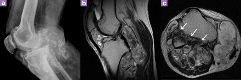
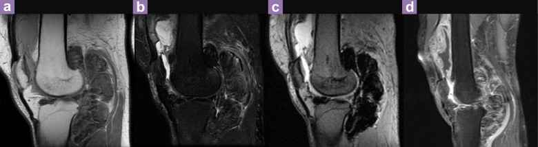
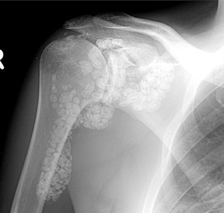
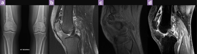
Definitive determination of the cause of articular swelling may be difficult based on just the clinical symptoms, physical examinations and laboratory tests. Joint disorders fall under the realms of rheumatology and general orthopaedics; however, patients with joint conditions manifesting primarily as intra-articular and peri-articular soft tissue swelling may at times be referred to an orthopaedic oncology department with suspicion of a tumour. In such a situation, an onco-radiologist needs to think beyond the usual neoplastic lesions and consider the diagnoses of various non-neoplastic arthritic conditions that may be clinically masquerading as masses. Differential diagnoses of articular lesions include infectious and non-infectious synovial proliferative processes, degenerative lesions, deposition diseases, vascular malformations, benign and malignant neoplasms and additional miscellaneous conditions. Many of these diseases have specific imaging findings. Knowledge of these radiological characteristics in an appropriate clinical context will allow for a more confident diagnosis.

Keywords: intra-articular; peri-articular; synovial; tumours.


Figures

Figure 1
Figure 1
Antero-posterior and lateral radiograph (a,b) of the right knee in a patient with parosteal osteosarcoma who presented with painful knee swelling and restriction of movement.
Figure 2
Figure 2
Chondrolipoma of the knee. A 29-year-old male patient presented with a painless, non-tender swelling on the lateral aspect of the left knee. Axial T2- weighted (a), coronal T1-weighted (b) and fat suppressed proton density (PD) weighted (c) images show a well-defined pear shaped fat signal intensity lesion with multiple internal curvilinear hypointensities deep to the lateral patellar retinaculum and extending beneath vastus lateralis muscle consistent with an atypical lipomatous lesion. Biopsy and histopathologic examination of the surgical specimen demonstrated a chondrolipoma.
Figure 3
Figure 3
A 16-year-old boy presented with an indolent swelling on the posterior aspect of the right knee. Lateral knee radiograph (a) reveals an ill-defined soft tissue density mass in the popliteal fossa with no obvious calcifications. Axial T1 and T2-weighted magnetic resonance images (b and c respectively) show a heterogeneous, multilobulated juxta-articular mass in the popliteal fossa (arrowheads) with septations and cystic areas. One of the lobules demonstrates hyper intense signal on T1 and fluid levels on T2-weighted image (arrows) compatible with hemorrhage. Histopathology examination demonstrated a biphasic synovial sarcoma.
Figure 4
Figure 4
A 54-year-old man with disseminated colon cancer presented with a focal, hard swelling on the medial aspect of left knee. Axial T1, T2 and fat suppressed PD-weighted image (a, b and c respectively) show a lobulated soft tissue mass in medial patello-femoral compartment of the left knee, involving medial patellar retinaculum and causing erosion of medial border of patella. The mass shows iso intense signal to muscle on T1 and hyper intense signal intensity on T2 and fat suppressed PD-weighted images. Biopsy examination of the lesion demonstrated adenocarcinoma with histology identical to the primary lesion in colon.
Figure 5
Figure 5
Synovial chondrosarcoma in a 71-year-old man presenting with a gradually progressive, mildly painful knee swelling and limitation of movement. Lateral radiographs of the right knee; (a) demonstrates an intra- and peri-articular mass with chondroid pattern of matrix mineralization. Sagittal and axial T2-weighted images, (b and c respectively) confirm the synovial origin of the mass with a predominant extra-articular component, extending into surrounding soft tissues and causing large bony erosions (arrows), features favoring an aggressive behavior. The mass shows a lobulated appearance with heterogeneously hyper intense signal intensity on T2-weighted images. Biopsy of the mass demonstrated a low grade chondrosarcoma.
Figure 6
Figure 6
A 34-year-old man with a large fluctuant right knee swelling. Sagittal T1 and fat suppressed T2-weighted images of the knee (a and b respectively) demonstrate a lobulated profoundly hypointense mass in the knee joint, predominantly along posterior aspect with evidence of magnetic susceptibility artifacts (blooming) on sagittal gradient image (c), consistent with the diagnosis of a pigmented villonodular synovitis which was confirmed on histopathology examination. Post-gadolinium T1-weighted image, (d) reveals intensely enhancing, marked nodular thickening of the synovium.
Figure 7
Figure 7
Synovial osteochondromatosis in a 40-year-old man presenting with right shoulder joint pain and restriction of movement. Anteroposterior radiograph of shoulder reveals innumerable, uniform sized, ossified intra-articular loose bodies at the glenohumeral joint and its synovial recesses. Note is made of preserved joint space and absence of significant degenerative joint changes.
Figure 8
Figure 8
Synovial chondromatosis in a patient with bilateral knee pain. Frontal radiograph of both knee joints (a) reveals multiple similar sized intra-articular loose bodies. Sagittal PD (b), gradient (c), and fat suppressed T2-weighted images (d) reveal synovial proliferation with intra-articular calcified and ossified loose bodies demonstrating uniformly hypointense signal and central marrow signal intensity with well corticated profoundly hypointense margins respectively.
Figure 9
Figure 9
Lipoma arborescence in a 46-year-old man with recurrent knee joint swelling and pain. Sagittal T1 (a) and fat suppressed T2-weighted (b) images of the knee joint reveal mild synovial effusion and prominent subsynovial fat along with frond-like projections into the synovial space of suprapatellar bursa, showing hyperintense signal on T1W image and signal loss on fat suppressed image compatible with fat containing villi.
Figure 10
Figure 10
Tuberculous arthritis. Axial fat suppressed post gadolinium T1-weighted image of the knee joint reveals markedly thickened and enhancing synovium (arrows) with effusion and loculations. Abbreviations: P = patella, F = femur.
Figure 11
Figure 11
Known case of chronic tophaceous gout presenting with left ankle swelling. Frontal radiograph of the left ankle (a) reveals soft tissue swelling (asterisk) in medial submalleolar region with well-defined erosion (arrow) of the tip of medial malleolus. Soft tissue fullness is also noted on the lateral aspect. Coronal T1 (b), T2-weighted (c) and axial gradient echo (d) magnetic resonance images demonstrate juxta-articular erosions of medial and lateral malleolus and talus (arrowheads in d) with preserved joint space and associated eccentric intra-articular tophaceous deposits (arrows) showing characteristic heterogeneous low signal intensity on T1 and T2-weighted images with absence of “blooming” on gradient image.
Figure 12
Figure 12
Biopsy-proven dialysis related amyloidosis in a 70-year-old man on hemodialysis for 21 years for diabetic nephropathy. Coronal computed tomography (CT) image of abdomen (a) reveals end stage renal disease with severe atherosclerotic vascular calcification. Axial CT image at the level of hip joints (b) demonstrates well-defined cystic lesions with sclerotic rim in both femoral heads and right femoral neck communicating with the joint space and intra-articular amyloid deposits.
Figure 13
Figure 13
Known case of rheumatoid arthritis with rice bodies. Lateral radiograph of the ankle (a) reveals a lobulated soft tissue swelling on its anterior aspect. Sagittal T1 and T2-weighted images (b and c) demonstrate fluid distension of the extensor digitorum longus tendon sheath (arrows) with intact tendon. Post-gadolinium fat suppressed T1-weighted image (d) reveals synovial thickening and intense enhancement of the tendon sheath. Features are consistent with tenosynovitis. Numerous uniform sized, tiny miliary T2 hypointense nodules noted within the synovial fluid are suggestive of rice bodies.
Figure 14
Figure 14
A 17-year-old boy with a palpable lump on the posterior aspect of the left knee. Lateral knee radiograph (a) reveals a soft tissue mass with bizzare calcification on its posterior aspect. Sagittal STIR MR image (b) shows a juxta-articular hyperintense mass with multiple round well-defined hypointense foci within suggestive of phleboliths. Axial post-gadolinium T1-weighted image (c) demonstrates avid contrast enhancement within the mass with infiltrative margins. Coronal T1-weighted image reveals few hyperintense foci within the mass (arrows). Biopsy demonstrated a synovial hemangioma.
Figure 15
Figure 15
A 42-year-old man with type I Arnold Chiari malformation, now presenting with a painless right shoulder mass. Sagittal T2-weighted image (d) of the cervical spine demonstrates tonsillar herniation (arrowhead) with a benign syrinx in upper cervical and upper thoracic cord (arrows). Antero-posterior radiograph of the right shoulder joint (a) reveals resorption of the upper end of humerus resembling surgical amputation associated with joint disorganization and large soft tissue swelling containing calcareous debris (arrows). Axial T1-weighted (b) and coronal fat suppressed T2-weighted (c) images confirm the radiographic findings (arrowhead = resorbed humerus) and reveal a large joint effusion (arrows). Aspiration of shoulder joint with a thick needle showed thick hemorrhagic aspirate with calcareous material which was negative for micro-organisms on staining and cultures. In the given clinical context, the imaging features are compatible with neuropathic joint.
Figure 16
Figure 16
Axial gradient echo image of knee joint in a known case of hemophilia reveals synovial thickening with profound signal hypointensity (arrowheads) suggestive of hemosiderin deposition from recurrent intra-articular bleeds.
Figure 17
Figure 17
Lipohemarthrosis in a patient with twisting knee injury. Lateral radiograph of the knee shows a fat-fluid level (arrowheads). Coronal T1-weighted image (b) reveals an intra-articular lateral tibial plateau fracture (arrow). Sagittal fat suppressed T2-weighted image (c) demonstrates three layers showing hematocrit effect (black arrow) and non-dependent fat layer (asterisk).
Figure 18
Figure 18
Diagnostic approach in patients presenting with suspected tumour-like swelling in and around joints.

Similar articles

Cited by

References

    1. Llauger J, Palmer J, Rosón N, Bagué S, Camins A, Cremades R. Nonseptic Monoarthritis: Imaging Features with Clinical and Histopathologic Correlation. Radiographics. 2000;20:S263–S278. - PubMed
    1. Sheldon PJ, Forrester DM, Learch TJ. Imaging of Intraarticular Masses. Radiographics. 2005;25(1):105–119. - PubMed
    1. Bredella MA, Stoller DW, Johnston JO. Magnetic Resonance Imaging in Orthopedics and sports medicine. In: Stoller DW, editor. Bone and Soft tissue tumors. 3rd ed. Philadelphia (US): Lippincott Williams & Wilkins; 2007. pp. 2045–2161.
    1. Lee FY, Keel SB, Gebhardt MC, Rosenthal DI. Intraarticular lipoma with osteochondroid metaplasia in the knee joint. Skeletal Radiol. 2001;30(4):230–233. - PubMed
    1. Marui T, Yamamoto T, Kimura T, Akisue T, Nagira K, Nakatani T, et al. A true intra-articular lipoma of the knee in a girl. Arthroscopy. 2002;18(5):E24. - PubMed

LinkOut - more resources