

MAS-mediated antioxidant effects restore the functionality of angiotensin converting enzyme 2-angiotensin-(1-7)-MAS axis in diabetic rat carotid
- PMID: 24877125
- PMCID: PMC4022170
- DOI: 10.1155/2014/640329
MAS-mediated antioxidant effects restore the functionality of angiotensin converting enzyme 2-angiotensin-(1-7)-MAS axis in diabetic rat carotid
Abstract
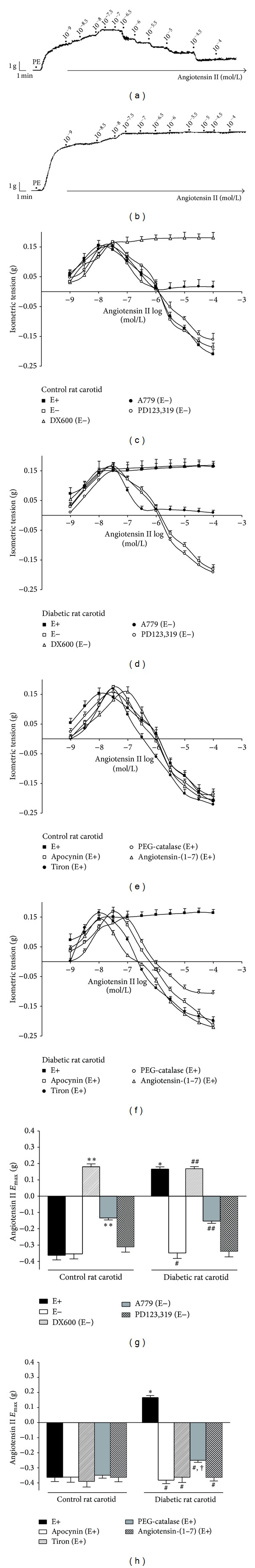
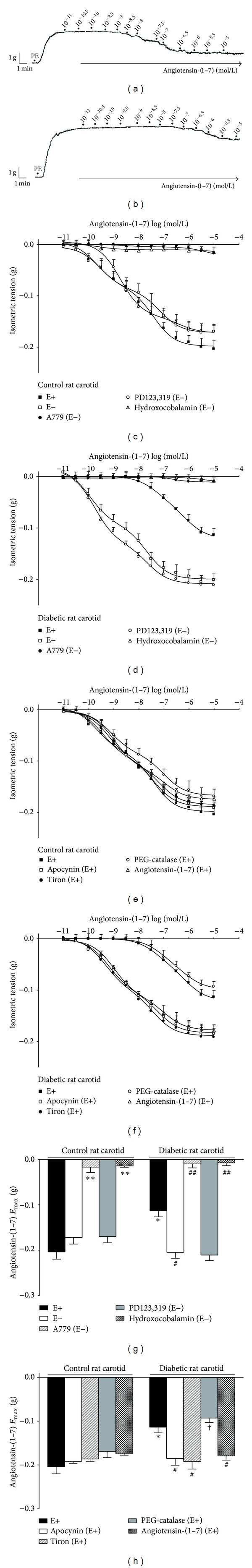
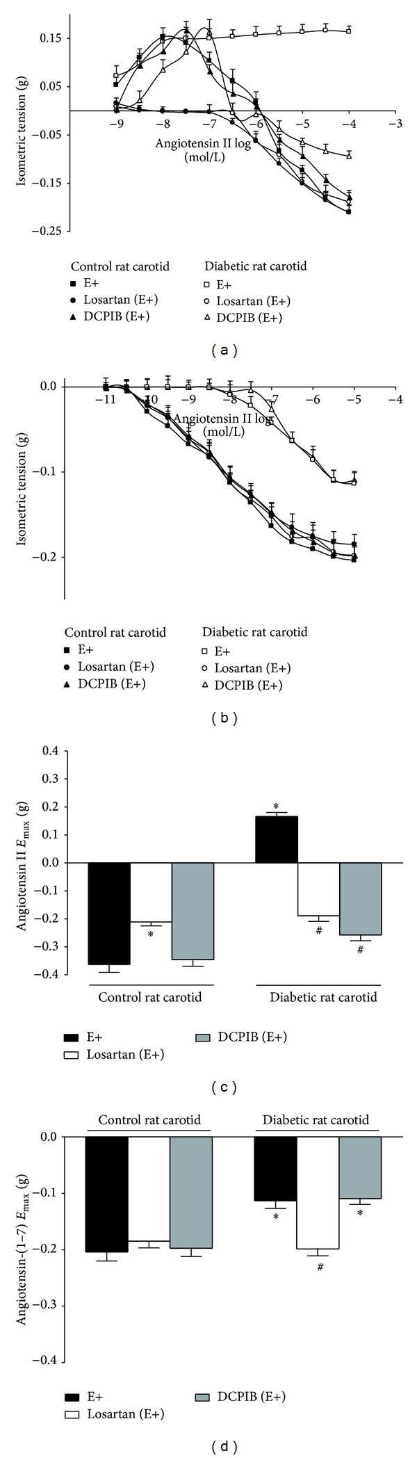
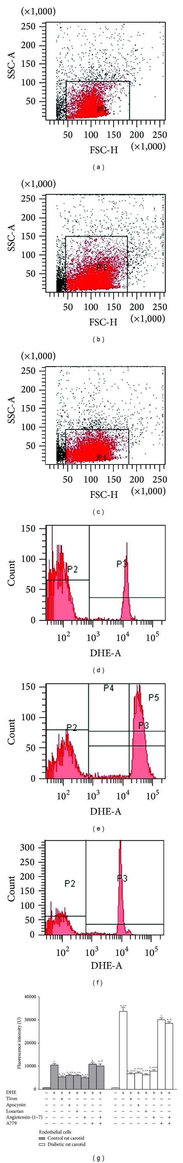
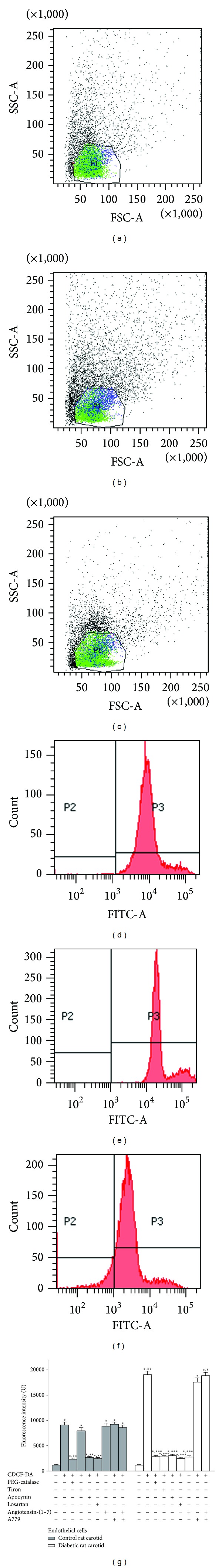
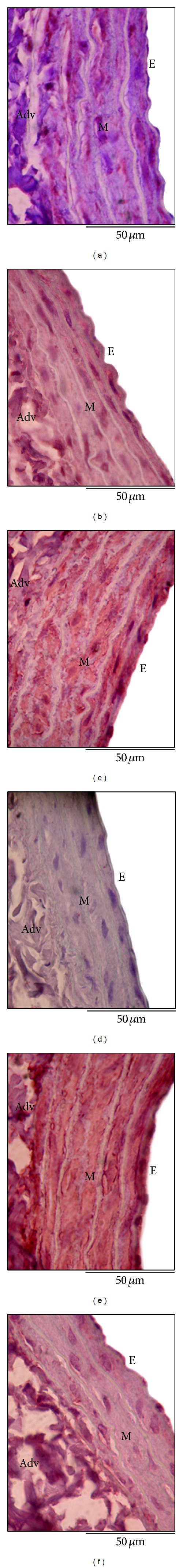
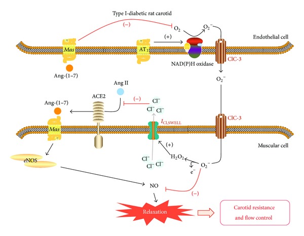
We hypothesized that endothelial AT1-activated NAD(P)H oxidase-driven generation of reactive oxygen species during type I-diabetes impairs carotid ACE2-angiotensin-(1-7)-Mas axis functionality, which accounts for the impaired carotid flow in diabetic rats. We also hypothesized that angiotensin-(1-7) chronic treatment of diabetic rats restores carotid ACE2-angiotensin-(1-7)-Mas axis functionality and carotid flow. Relaxant curves for angiotensin II or angiotensin-(1-7) were obtained in carotid from streptozotocin-induced diabetic rats. Superoxide or hydrogen peroxide levels were measured by flow cytometry in carotid endothelial cells. Carotid flow was also determined. We found that endothelial AT1-activated NAD(P)H oxidase-driven generation of superoxide and hydrogen peroxide in diabetic rat carotid impairs ACE2-angiotensin-(1-7)-Mas axis functionality, which reduces carotid flow. In this mechanism, hydrogen peroxide derived from superoxide dismutation inhibits ACE2 activity in generating angiotensin-(1-7) seemingly by activating I(Cl,SWELL0, while superoxide inhibits the nitrergic Mas-mediated vasorelaxation evoked by angiotensin-(1-7). Angiotensin-(1-7) treatment of diabetic rats restored carotid ACE2-angiotensin-(1-7)-Mas axis functionality by triggering a positive feedback played by endothelial Mas receptors, that blunts endothelial AT1-activated NAD(P)H oxidase-driven generation of reactive oxygen species. Mas-mediated antioxidant effects also restored diabetic rat carotid flow, pointing to the contribution of ACE2-angiotensin-(1-7)-Mas axis in maintaining carotid flow.
Figures
Similar articles
-
MAS receptors mediate vasoprotective and atheroprotective effects of candesartan upon the recovery of vascular angiotensin-converting enzyme 2-angiotensin-(1-7)-MAS axis functionality.Eur J Pharmacol. 2015 Oct 5;764:173-188. doi: 10.1016/j.ejphar.2015.07.007. Epub 2015 Jul 2. Eur J Pharmacol. 2015. PMID: 26144375
-
[Effect of Chinese herbs for stasis removing and collaterals dredging upon angiotensin-converting enzyme 2-angiotensin-(1-7)-mas axis in the renal cortex of diabetic nephropathy rats].Zhongguo Zhong Xi Yi Jie He Za Zhi. 2014 Jun;34(6):714-21. Zhongguo Zhong Xi Yi Jie He Za Zhi. 2014. PMID: 25046956 Chinese.
-
Upregulation of ACE2-ANG-(1-7)-Mas axis in jejunal enterocytes of type 1 diabetic rats: implications for glucose transport.Am J Physiol Endocrinol Metab. 2012 Sep 1;303(5):E669-81. doi: 10.1152/ajpendo.00562.2011. Epub 2012 Jul 17. Am J Physiol Endocrinol Metab. 2012. PMID: 22811473
-
The Anti-Inflammatory Potential of ACE2/Angiotensin-(1-7)/Mas Receptor Axis: Evidence from Basic and Clinical Research.Curr Drug Targets. 2017;18(11):1301-1313. doi: 10.2174/1389450117666160727142401. Curr Drug Targets. 2017. PMID: 27469342 Review.
-
ACE2, angiotensin-(1–7), and Mas: the other side of the coin.Pflugers Arch. 2013 Jan;465(1):79-85. doi: 10.1007/s00424-012-1120-0. Pflugers Arch. 2013. PMID: 23463883 Review.
Cited by
-
Angiotensin-(1-7) as a Potential Therapeutic Strategy for Delayed Cerebral Ischemia in Subarachnoid Hemorrhage.Front Immunol. 2022 Mar 9;13:841692. doi: 10.3389/fimmu.2022.841692. eCollection 2022. Front Immunol. 2022. PMID: 35355989 Free PMC article. Review.
-
Effects of renal denervation on cardiac oxidative stress and local activity of the sympathetic nervous system and renin-angiotensin system in acute myocardial infracted dogs.BMC Cardiovasc Disord. 2017 Feb 17;17(1):65. doi: 10.1186/s12872-017-0498-1. BMC Cardiovasc Disord. 2017. PMID: 28212603 Free PMC article.
-
The Impact of Angiotensin-Converting Enzyme-2/Angiotensin 1-7 Axis in Establishing Severe COVID-19 Consequences.Pharmaceutics. 2022 Sep 8;14(9):1906. doi: 10.3390/pharmaceutics14091906. Pharmaceutics. 2022. PMID: 36145655 Free PMC article. Review.
-
The ACE2/Angiotensin-(1-7)/MAS Axis of the Renin-Angiotensin System: Focus on Angiotensin-(1-7).Physiol Rev. 2018 Jan 1;98(1):505-553. doi: 10.1152/physrev.00023.2016. Physiol Rev. 2018. PMID: 29351514 Free PMC article. Review.
-
Vascular endothelial dysfunction and pharmacological treatment.World J Cardiol. 2015 Nov 26;7(11):719-41. doi: 10.4330/wjc.v7.i11.719. World J Cardiol. 2015. PMID: 26635921 Free PMC article. Review.
References
-
- Glowinska-Olszewska B, Hyniewicz A, Jeznach M, et al. Relationship between circulating endothelial progenitor cells and endothelial dysfunction in children with type 1 diabetes: a novel paradigm of early atherosclerosis in high-risk young patients. European Journal of Endocrinology. 2013;168:153–161. - PubMed
-
- Hurks R, Eisinger MJ, Goovaerts I, et al. Early endothelial dysfunction in young type 1 diabetics. European Journal of Vascular and Endovascular Surgery. 2009;37(5):611–615. - PubMed
-
- Yousif MHM, Benter IF, Akhtar S. The role of tyrosine kinase-mediated pathways in diabetes-induced alterations in responsiveness of rat carotid artery. Autonomic and Autacoid Pharmacology. 2005;25(2):69–78. - PubMed
Publication types
MeSH terms
Substances
LinkOut - more resources
Full Text Sources
Other Literature Sources
Medical
Research Materials
