. 2014:2014:691041.
doi: 10.1155/2014/691041. Epub 2014 Apr 27.

Inflammatory profiling of Schwann cells in contact with growing axons distal to nerve injury

Affiliations

Inflammatory profiling of Schwann cells in contact with growing axons distal to nerve injury

Petr Dubový et al. Biomed Res Int. 2014.

Abstract

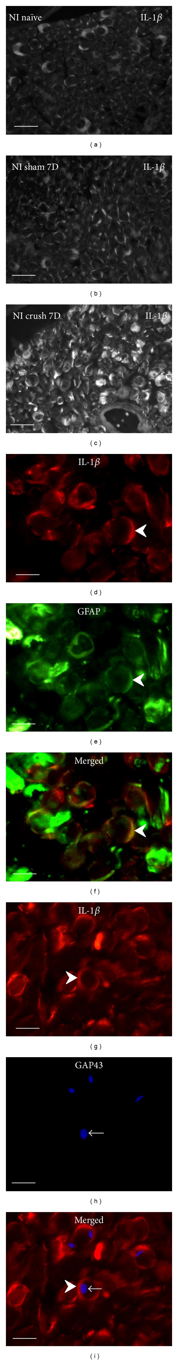
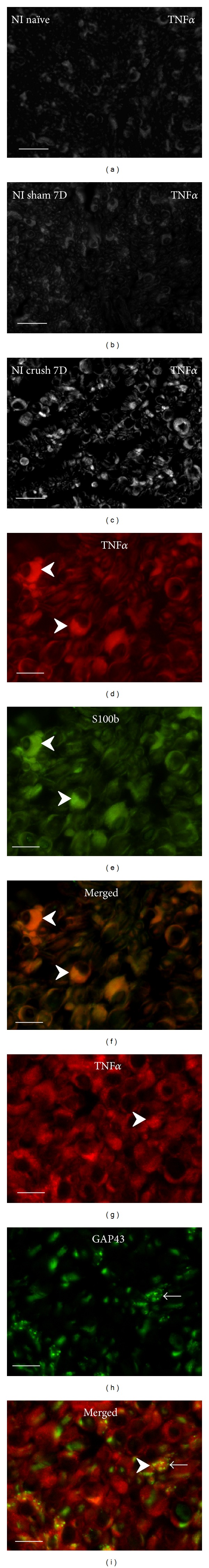
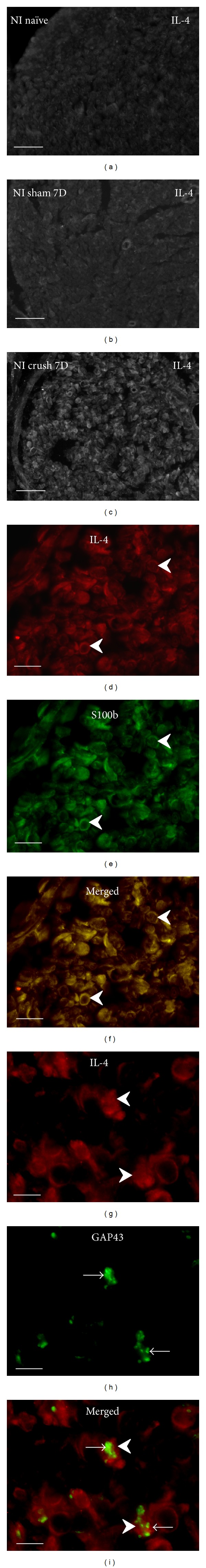
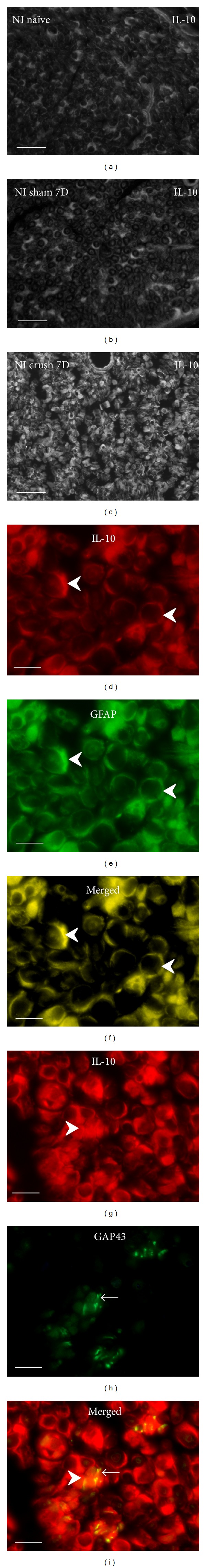
Activated Schwann cells distal to nerve injury upregulate inflammatory mediators, including cytokines. The goal of the present study was to investigate expression of proinflammatory (IL-1β, TNFα) and anti-inflammatory cytokines (IL-4, IL-10) in activated Schwann cells in relation to growing axons distal to crush injury of rat sciatic nerves. Seven days from sciatic nerve crush, transverse cryostat sections were cut 5 mm distal to lesion and incubated for double immunostaining to indicate Schwann cells (GFAP or S100b) and individual investigated cytokines or to demonstrate growing axons (GAP43). The Schwann cells of naïve sciatic nerves and those removed from sham-operated rats displayed similar weak immunoreactivity for the investigated cytokines. In contrast, increased intensity of cytokine immunofluorescence was found in Schwann cells distal to crush lesion. The cytokine-positive Schwann cells were found in close contact with growing axons detected by immunostaining for GAP43. The results of immunohistochemical analysis distal to nerve crush injury suggest that inflammatory profiling of Schwann cells including upregulation of both pro- and anti-inflammatory cytokines does not prevent growth of axons distal to nerve crush injury.


Figures

Figure 1
Figure 1
Cryostat cross sections through naïve rat sciatic nerve (a), after sham operation (b), and distal to crush for 7 d (c) immunostained for IL-1β revealed robust increase of immunofluorescence distal to nerve injury. Increased IL-1β immunofluorescence was found dominantly in Schwann cells detected by colocalization with GFAP (arrowheads, (d)–(f)). Growing axons and sprouts immunostained with GAP43 (arrows) were found in close contact with Schwann cells decorated by IL-1β immunostaining (arrowheads) ((g)–(i)). Scale bars for (a)–(c) = 20 μm, for (d)–(i) = 5 μm.
Figure 2
Figure 2
Representative cryostat sections through naïve rat sciatic nerve (a), after sham operation (b), and distal to crush for 7 d (c) immunostained for TNFα demonstrated strong increase of immunofluorescence intensity distal to nerve crush. Immunofluorescence staining for TNFα protein was observed in Schwann cells detected by colocalization with S100b immunostaining (arrowheads, (d)–(f)). Many growing axons immunostained for GAP43 (arrows) were closely related to Schwann cells decorated by TNFα immunostaining (arrowheads) ((g)–(i)). Scale bars for (a)–(c) = 80 μm, for (d)–(i) = 10 μm.
Figure 3
Figure 3
Cryostat sections through the sciatic nerve of naïve rat (a), after sham operation (b), and distal to sciatic nerve crush for 7 d (c) immunostained for IL-4. The sections of naive and sham-operated sciatic nerve displayed very weak immunofluorescence for IL-4, but moderate intensity of IL-4 immunofluorescence was observed distal to nerve crush injury. Schwann cells immunodetected by GFAP displayed also immunostaining for IL-4 at higher power magnification (arrowheads, (d)–(f)). Growing GAP43+ axons (arrows) were found in close contact with IL4+ Schwann cells (arrowheads) ((g)–(i)). Scale bars for (a)–(c) = 80 μm, for (d)–(f) = 15 μm, for (g)–(i) = 5 μm.
Figure 4
Figure 4
Cryostat sections through naïve rat sciatic nerve (a), after sham operation (b), and distal to crush for 7 d (c) illustrate robust increase of IL-10 immunofluorescence distal to nerve crush injury. Increased IL-10 immunofluorescence was found in GFAP+ Schwann cells (arrowheads, (d)–(f)). Many growing axons immunostained for GAP43 (arrows) were in close contact with Schwann cells immunopositive for IL-10 (arrowheads) ((g)–(i)). Scale bars for (a)–(c) = 40 μm, for (d)–(i) = 10 μm.

References

    1. Borthwick LA, Wynn TA, Fisher AJ. Cytokine mediated tissue fibrosis. Biochimica Et Biophysica Acta-Molecular Basis of Disease. 2013;1832(7):1049–1060. - PMC - PubMed
    1. Gaudet AD, Popovich PG, Ramer MS. Wallerian degeneration: gaining perspective on inflammatory events after peripheral nerve injury. Journal of Neuroinflammation. 2011;8, article 110 - PMC - PubMed
    1. Gensel JC, Kigerl KA, Mandrekar-Colucci SS, Gaudet AD, Popovich PG. Achieving CNS axon regeneration by manipulating convergent neuro-immune signaling. Cell and Tissue Research. 2012;349(1):201–213. - PMC - PubMed
    1. Moalem G, Tracey DJ. Immune and inflammatory mechanisms in neuropathic pain. Brain Research Reviews. 2006;51(2):240–264. - PubMed
    1. Kerschensteiner M, Meinl E, Hohlfeld R. Neuro-immune crosstalk in CNS diseases. Neuroscience. 2009;158(3):1122–1132. - PubMed

Publication types

LinkOut - more resources