. 2014 Jun;142(2):176-92.
doi: 10.1111/imm.12229.

Regulation of T-lymphocyte motility, adhesion and de-adhesion by a cell surface mechanism directed by low density lipoprotein receptor-related protein 1 and endogenous thrombospondin-1

Regulation of T-lymphocyte motility, adhesion and de-adhesion by a cell surface mechanism directed by low density lipoprotein receptor-related protein 1 and endogenous thrombospondin-1

Toomas Talme et al. Immunology. 2014 Jun.

Abstract

T lymphocytes are highly motile and constantly reposition themselves between a free-floating vascular state, transient adhesion and migration in tissues. The regulation behind this unique dynamic behaviour remains unclear. Here we show that T cells have a cell surface mechanism for integrated regulation of motility and adhesion and that integrin ligands and CXCL12/SDF-1 influence motility and adhesion through this mechanism. Targeting cell surface-expressed low-density lipoprotein receptor-related protein 1 (LRP1) with an antibody, or blocking transport of LRP1 to the cell surface, perturbed the cell surface distribution of endogenous thrombospondin-1 (TSP-1) while inhibiting motility and potentiating cytoplasmic spreading on intercellular adhesion molecule 1 (ICAM-1) and fibronectin. Integrin ligands and CXCL12 stimulated motility and enhanced cell surface expression of LRP1, intact TSP-1 and a 130,000 MW TSP-1 fragment while preventing formation of a de-adhesion-coupled 110 000 MW TSP-1 fragment. The appearance of the 130 000 MW TSP-1 fragment was inhibited by the antibody that targeted LRP1 expression, inhibited motility and enhanced spreading. The TSP-1 binding site in the LRP1-associated protein, calreticulin, stimulated adhesion to ICAM-1 through intact TSP-1 and CD47. Shear flow enhanced cell surface expression of intact TSP-1. Hence, chemokines and integrin ligands up-regulate a dominant motogenic pathway through LRP1 and TSP-1 cleavage and activate an associated adhesion pathway through the LRP1-calreticulin complex, intact TSP-1 and CD47. This regulation of T-cell motility and adhesion makes pro-adhesive stimuli favour motile responses, which may explain why T cells prioritize movement before permanent adhesion.


Figures

Figure 1
Figure 1
Ustekinumab inhibits T-lymphocyte motility. (a) Photographs showing the morphology of cells in a collagen gel after incubation in medium only or in medium containing 100 μg/ml of ustekinumab or infliximab for 15 min. (b) Migration of cells into a collagen gel at different levels after incubation in the presence of ustekinumab (100 μg/ml) or infliximab (100 μg/ml) for 30 min. (c) Migration of cells into a collagen gel at different levels after incubation in the presence of ustekinumab (100 μg/ml) for 30 min and subsequently in the absence of ustekinumab for 2 hr before determination of migration. Removal of ustekinumab restored polarized cell shape and motility showing that the inhibitory effect was reversible. (d) The influence of ustekinumab (100 μg/ml) or infliximab (100 μg/ml) on transendothelial migration of the T-cell clone AF24 in a flow system using confluent tumour necrosis factor-pre-treated endothelial cells overlaid with CXCL12 10 ng/ml and the subendothelial compartment filled with medium containing CCL5 100 ng/ml. The ordinates show the accumulated number of transmigrated cells from six passages. The results in (b) and (c) show mean values of three independent experiments and (d) shows mean values of two independent experiments.
Figure 2
Figure 2
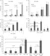
Ustekinumab inhibits T-lymphocyte motility through lipoprotein receptor-related protein 1 (LRP1) and thrombospondin 1 (TSP-1). (a,b) Motility was examined with cells in the presence of ustekinumab (100 μg/ml) or infliximab (100 μg/ml on intercellular adhesion molecule 1 (10 μg/ml) (a) and fibronectin (10 μg/ml) (b) as determined by a transwell assay measuring number of transmigrated cells. (c) Formation of conjugates of AF24 T cells with antigen-presenting HLA-identical B cells in the presence of ustekinumab (100 μg/ml) or infliximab (100 μg/ml). (d) Gel analysis (6% SDS–PAGE) showing the influence of incubation with ustekinumab on the cell surface expression of TSP-1, LRP1 and CD4 after 15 min, as demonstrated by immunoprecipitation of surface biotinylated cells. (e) Quantitative immunocytochemistry showing the influence of ustekinumab (100 μg/ml) and infliximab (100 μg/ml) on cell-surface-expressed and intracellular LRP1 and TSP-1 as determined after 30 min. The cell surface expression of CD4 is shown as a comparison. (a,b) Mean values of three independent experiments and (c) one representative experiment of two independent experiments. The results in (d) and (e) show one representative experiment of three to five independent experiments.
Figure 3
Figure 3
The influence of various inhibitors on the cell surface expression of thrombospondin 1 (TSP-1) and lipoprotein receptor-related protein 1 (LRP1) and on T-cell motility. (a,b) Cell surface expression of TSP-1 (a) and LRP1 (b) after incubation with ustekinumab (100 μg/ml) alone, with ustekinumab plus cycloheximide (10 μg/ml), dynasore (80 μm), colchicine (10−6 m) and brefeldin A (10 μg/ml) or with inhibitors only for 30 min. (c) The influence of dynasore, colchicine and ustekinumab [same concentrations as in (a,b)] on T-cell motility measured as total number of migrated cells at different levels throughout a collagen gel after 30 min. (d) Dynasore and ustekinumab induce [same concentrations as in (a,b)] redistribution of TSP-1 on the cell surface expressed as percentage cells with a cap-like distribution. The cells were incubated with inhibitors for 15 min before fixation. (e) Photographs of cells showing capping of TSP-1 after incubation with dynasore and ustekinumab or dynasore plus ustekinumab for 15 min. The results in (a–c) show mean values of three independent experiments. The results in (d) and (e) show one representative experiment of three independent experiments.
Figure 4
Figure 4
Ustekinumab inhibits CXCL12-induced T-lymphocyte motility and effects on lipoprotein receptor-related protein 1 (LRP1) and thrombospondin 1 (TSP-1). (a) The influence of ustekinumab (100 μg/ml) on migration into a collagen matrix in the absence and presence of CXCL12 (50 ng/ml) as determined after 30 min. (b) Gel analysis [4–12% SDS–PAGE (LRP1) and 6% SDS–PAGE (TSP-1)] showing the influence of ustekinumab, CXCL12 and dynasore on the cell surface expression of TSP-1 and LRP1. The cells were surface biotinylated and immunoprecipitated after 10 min. (c) Western blotting (6% SDS–PAGE) of material from lysed cells after incubation for 30 min with and without CXCL12 (50 ng/ml). (d) Western blotting of material from lysed cells using different anti-TSP-1 antibodies (Ab9 reacts with the N-terminal domain of TSP-1) after incubation for 30 min with CXCL12 (50 ng/ml). The results in (a) show mean values of three independent experiments. The results in (b–d) show one representative experiment of three to seven independent experiments.
Figure 5
Figure 5
Cell surface expression of lipoprotein receptor-related protein 1 (LRP1) and thrombospondin 1 (TSP-1) in adherent and de-adherent cells, and the influence of ustekinumab on adhesion. (a) Gel analysis showing the surface expression of LRP1, TSP-1 (Ab3), CD4 and CD29 in cells in suspension and after adhesion to fibronectin (10 μg/ml) and intercellular adhesion molecule 1 (ICAM-1; 2 μg/ml) for 30 min before biotinylation and immunoprecipitation. (b) Gel analysis as described in Fig. 4(a) showing the cell surface expression of LRP1, TSP-1 and CD29 in cells allowed to adhere to ICAM-1 for 30 min. The substrata coated with fibronectin and ICAM-1 were then washed to remove non-adherent cells and allowed to incubate for 20 min. Cells that had detached during this 20-min incubation were collected by aspiration of the medium and gentle washing. Subsequently, adherent and de-adherent cells were biotinylated, collected and immunoprecipitated. It is evident that de-adhesion reverses cell surface expression of intact TSP-1 and 130 000 MW TSP-1 (Ab3) and LRP1 and induces a 110 000 MW band reactive with anti-TSP-1 antibodies. (c) Influence of ustekinumab and infliximab on cell shape, measured as per cent polarized cells, after adhesion to ICAM-1 (2 μg/ml) for 10 min in the presence and absence of CXCL12. (d) Morphology of adherent cells in response to ustekinumab, infliximab and CXCL12. (a, b and d) Representative experiments of four to seven independent experiments and (c) shows mean values of three independent experiments.
Figure 6
Figure 6
Free-floating and adherent cells differ with respect to the cell surface expression of lipoprotein receptor-related protein 1 (LRP1) and thrombospondin 1 (TSP-1). Gel analysis showing the cell surface expression of TSP-1 and LRP1 in untreated control cells, free-floating cells and cells adherent to fibronectin (10 μg/ml) and ICAM-1 (2 μg/ml) for 15 min before biotinylation and immunoprecipitation. The results show one representative experiment of four independent experiments.
Figure 7
Figure 7
Differential roles of lipoprotein receptor-related protein 1 (LRP1 and thrombospondin 1 (TSP-1 in adhesion. (a,b) The influence of transfection with control siRNA (a) and LRP1 siRNA (b) on adhesion of T cells from healthy individuals to intercellular adhesion molecule 1 (ICAM-1; 2 μg/ml) as determined at different times under static conditions and when exposed to shear flow. (c,d) The influence of exogenous TSP-1 (c) and of exogenous TSP-1 and a peptide mimetic of the CD47 binding site in TSP-1 (4N1K) (d) on adhesion of AF24 T cells to ICAM-1 as determined after 30 min. AF24 were cultured in the presence of cycloheximide (10 μg/ml) for 4 hr before the experiment. Incubation with cycloheximide for 2 hr reduced the expression of TSP-1 significantly whereas incubation for 30 min did not affect the amount of TSP-1 in the cells (not shown). (e) The influence of a peptide mimetic of the TSP-1 binding site in calreticulin (CRT19–36), a scrambled control peptide, and intact exogenous TSP-1 on adhesion of cycloheximide-treated AF24 T cells to ICAM-1 as determined after 30 min. The results show one representative experiment of two to four independent experiments.
Figure 8
Figure 8
A mechanism for integrated regulation of motility and adhesion in T cells and its role for transendothelial migration. (a) CXCL12 and integrin ligands up-regulate the cell surface expression of thrombospondin 1 (TSP-1) and lipoprotein receptor-related protein 1 (LRP1). The LRP1 induces motogenic signalling through Janus kinase (JAK) whereas intact TSP-1 induces adhesion to intercellular adhesion molecule 1 (ICAM-1) and fibronectin through its C-terminal domain via CD47. This ‘adhesion pathway’ is triggered by interaction of LRP1-associated calreticulin with the N-terminal domain of TSP-1. Interaction of the N-terminal domain of TSP-1 with LRP1 concomitantly induces cleavage of TSP-1, which accelerates the dominant LRP1-directed motogenic pathway through a 130 000 MW TSP-1 fragment and inhibits the triggering of adhesion. Enhancement of the cell surface expression of intact TSP-1 by shear flow enhances the adhesion pathway. Processing of TSP-1 to a 110 000 MW fragment and reducing expression of LRP1 and associated calreticulin inhibits adhesion. Based on the findings in Fig. 3 TSP-1 is implied to appear as an independent component within the plasma membrane but, owing to limited space, TSP-1 is placed over the membrane. (b) The possible influence of cell surface-expressed TSP-1 and LRP1 induced by CXCL12 and integrin ligands on transendothelial migration. Free-floating cells up-regulate cell surface expression of intact TSP-1. After initial rolling, loosely attached cells in close proximity of the endothelium are activated by CXCL12, which induces cell surface expression of LRP1 and enhances TSP-1 expression. LRP1-calreticulin promotes firm adhesion/spreading to ICAM-1 through intact TSP-1 and CD47 and the association of CD47 to LFA-1. Adhesion and spreading on the endothelium is inhibited by TSP-1 cleavage. LRP1 and 130 000 MW TSP-1 stimulate migration through the endothelium via JAK signalling. Integrins may bypass the need for chemokine signals to initiate transendothelial migration. Subendothelial chemokines or chemokines in endothelial junctions stimulate diapedesis. LRP1-dependent TSP-1 cleavage makes the motogenic LRP1–JAK pathway dominate over the pro-adhesive TSP-1-CD47 pathway.

Similar articles

Cited by

References

    1. Christensen JE, Nansen A, Moos T, Lu B, Gerard C, Christensen JP, Thomsen AR. Efficient T-cell surveillance of the CNS requires expression of the CXC chemokine receptor 3. J Neurosci. 2004;24:4849–58. - PMC - PubMed
    1. Ousman SS, Kubes P. Immune surveillance in the central nervous system. Nat Neurosci. 2012;15:1096–101. - PMC - PubMed
    1. Schaerli P, Ebert L, Willimann K, Blaser A, Roos RS, Loetscher P, Moser B. A skin-selective homing mechanism for human immune surveillance T cells. J Exp Med. 2004;199:1265–75. - PMC - PubMed
    1. Swann JB, Smyth MJ. Immune surveillance of tumors. J Clin Invest. 2007;117:1137–46. - PMC - PubMed
    1. Meiron M, Zohar Y, Anunu R, Wildbaum G, Karin N. CXCL12 (SDF-1α) suppresses ongoing experimental autoimmune encephalomyelitis by selecting antigen-specific regulatory T cells. J Exp Med. 2008;205:2643–55. - PMC - PubMed

Publication types

MeSH terms

Substances