. 2014 Aug:125:9-19.
doi: 10.1016/j.exer.2014.05.014. Epub 2014 May 27.

Massive formation of square array junctions dramatically alters cell shape but does not cause lens opacity in the cav1-KO mice

Affiliations

Massive formation of square array junctions dramatically alters cell shape but does not cause lens opacity in the cav1-KO mice

Sondip K Biswas et al. Exp Eye Res. 2014 Aug.

Abstract

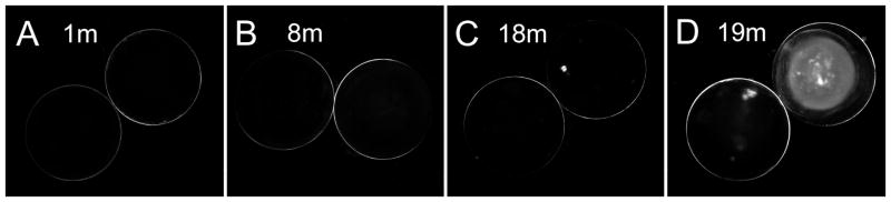
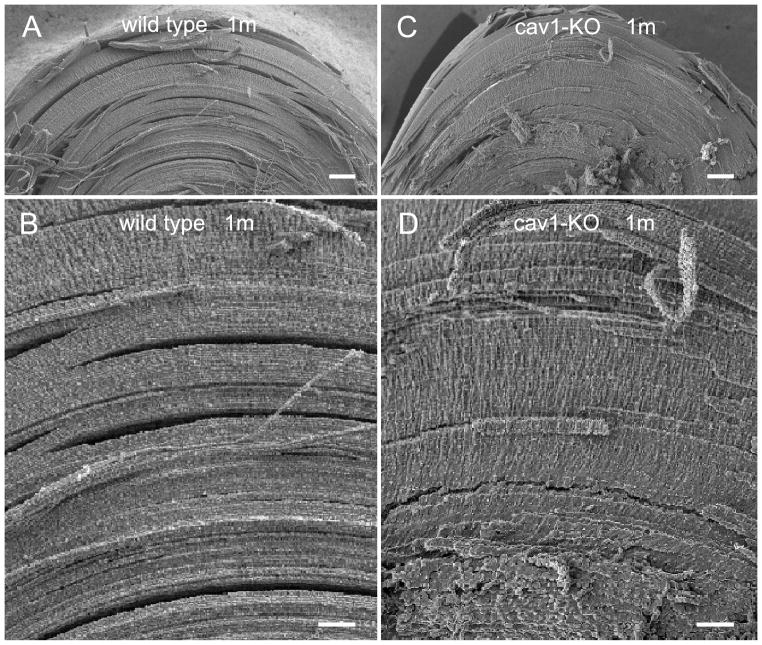
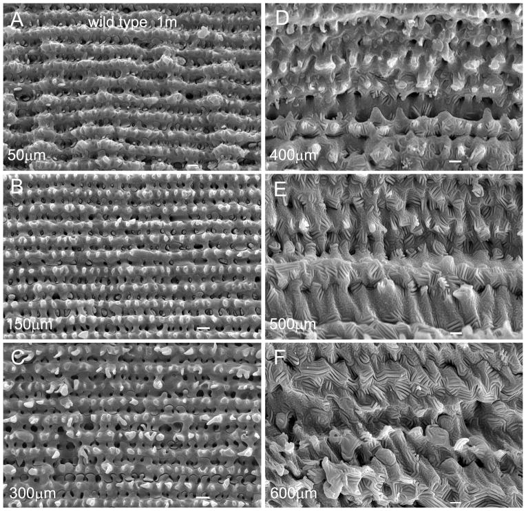
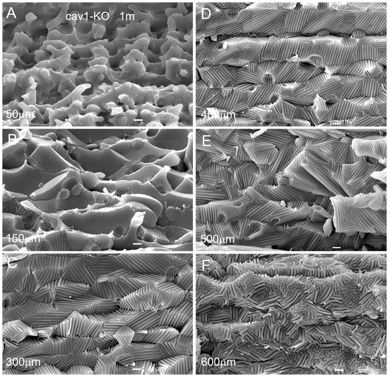
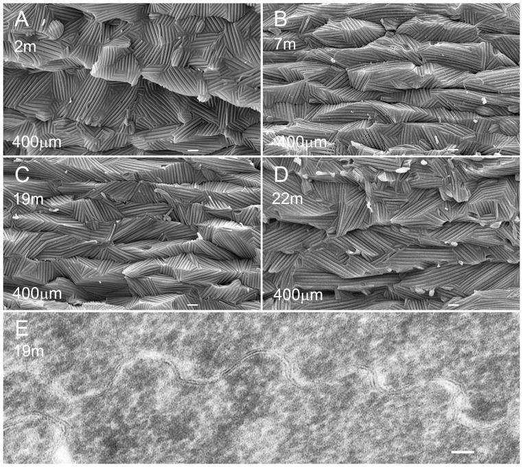
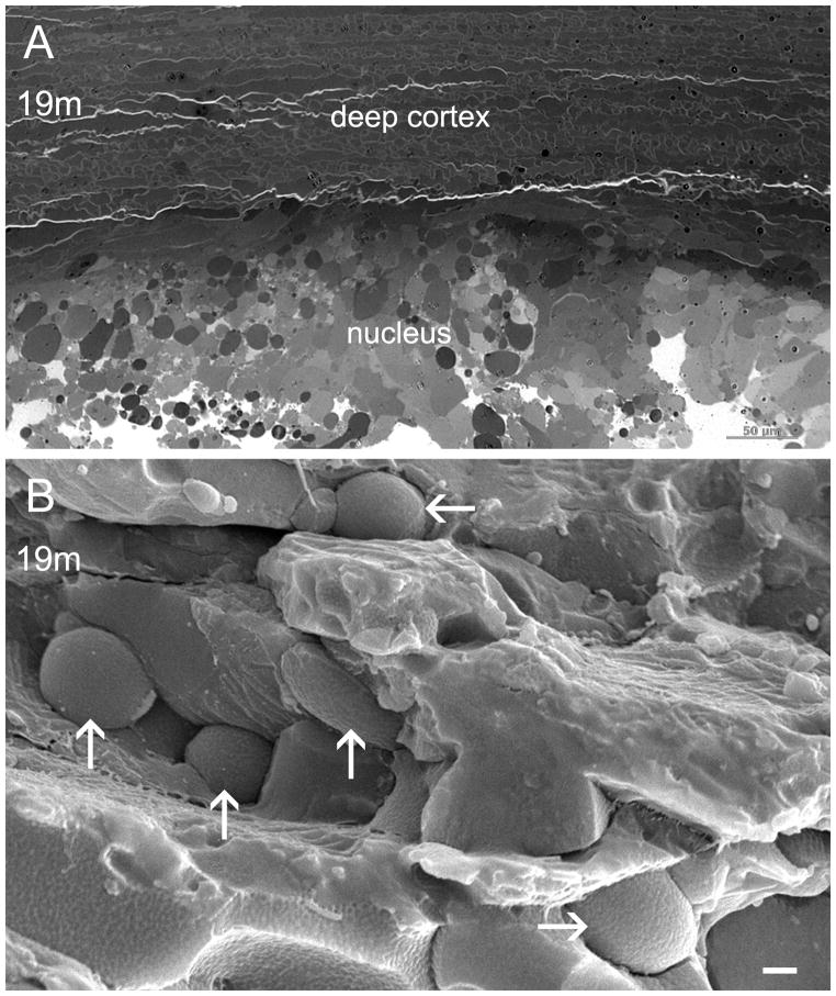
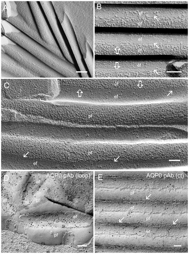
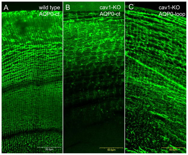
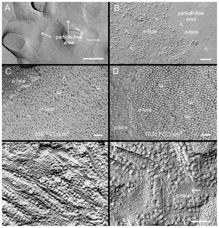
The wavy square array junctions are composed of truncated aquaporin-0 (AQP0) proteins typically distributed in the deep cortical and nuclear fibers in wild-type lenses. These junctions may help maintain the narrowed extracellular spaces between fiber cells to minimize light scattering. Herein, we investigate the impact of the cell shape changes, due to abnormal formation of extensive square array junctions, on the lens opacification in the caveolin-1 knockout mice. The cav1-KO and wild-type mice at age 1-22 months were used. By light microscopy examinations, cav1-KO lenses at age 1-18 months were transparent in both cortical and nuclear regions, whereas some lenses older than 18 months old exhibited nuclear cataracts. Scanning EM consistently observed the massive formation of ridge-and-valley membrane surfaces in young fibers at approximately 150 μm deep in all cav1-KO lenses studied. In contrast, the typical ridge-and-valleys were only seen in mature fibers deeper than 400 μm in wild-type lenses. The resulting extensive ridge-and-valleys dramatically altered the overall cell shape in cav1-KO lenses. Remarkably, despite dramatic shape changes, these deformed fiber cells remained intact and made close contact with their neighboring cells. By freeze-fracture TEM, ridge-and-valleys exhibited the typical orthogonal arrangement of 6.6 nm square array intramembrane particles and displayed the narrowed extracellular spaces. Immunofluorescence analysis showed that AQP0 C-terminus labeling was significantly decreased in outer cortical fibers in cav1-KO lenses. However, freeze-fracture immunogold labeling showed that the AQP0 C-terminus antibody was sparsely distributed on the wavy square array junctions, suggesting that the cleavage of AQP0 C-termini might not yet be complete. The cav1-KO lenses with nuclear cataracts showed complete cellular breakdown and large globule formation in the lens nucleus. This study suggests that despite dramatic cell shape changes, the massive formation of wavy square array junctions in intact fibers may provide additional adhesive support for maintaining the narrowed extracellular spaces that are crucial for the transparency of cav1-KO lenses.

Keywords: AQP0; caveolin-1 knockout; fiber cell membrane; lens; mouse; square array junctions.


Figures

Figure 1
Figure 1
Representative photographs of lens transparency in cav1-KO mice at different ages. All lenses examined in cav1-KO mice at age 1 to 18 months are transparent. A–C show transparent lenses at 1 month, 8 months and 18 months of age. Some lenses at 19 months and older display nuclear cataracts with various densities in the cav1-KO mice (D).
Figure 2
Figure 2
SEM comparison of intact cortical fibers in wild-type and cav1-KO lenses at low magnifications. The similarity in structural compactness between intact cortical fibers of wild-type (A and B) and cav1-KO (C and D) is discernible at the low magnification micrographs. Scale bars: A and C= 100 μm; B and D=50 μm.
Figure 3
Figure 3
Normal cell shape and membrane surface of cortical fibers in wild-type lenses. The cell membranes of normal cortical fiber cells change gradually from the smooth surface in the superficial cortex (A) to more irregular surface with elaborate interlocking protrusions in the outer cortex (B, C), approximately from 50 μm to 300 μm deep from the equatorial lens surface. The ridge-and-valley surface patterns, which represent wavy aquaporin junctions of fiber cells, are first seen in the inner cortex (D), approximately 400 μm deep from the lens surface. These ridge-and-valley surface patterns are found extended toward the entire deep cortical regions (i.e., 600 μm deep) of the lens (E, F). Scale bars: A–F = 1 μm.
Figure 4
Figure 4
Changes in cell shape and membrane surface of cortical fibers in cav1-KO lenses. SEM shows the extensive ridge-and-valley membrane surface patterns and dramatic alterations in cell shapes of cortical fiber cells in the cav1-KO lenses at 1 month old as compared with the age-matched wild-type. The dramatic changes of superficial fiber cells (e.g., approximately 50 μm deep) include swelling, irregular shape and smooth cell borders without interlocking protrusions (A). The irregular fiber cells begin to display small number and degree of ridge-and-valleys in outer cortex, approximately 150 μm deep from the lens surface (B). The membranes of fiber cells subsequently undergo a massive formation of extensive ridge-and-valley surface patterns extended toward the entire deep cortical regions (C–F), approximately 600 μm deep from the equatorial lens surface. These extensive ridge-and-valleys are regularly arranged into many elongated bundles with different orientations. They are closely interlocked with those of the neighboring cells, and thus resulting in forming tight intercellular spaces among all surrounding cortical fiber cells. Scale bars: A–F = 1 μm.
Figure 5
Figure 5
Similarity of ridge-and-valleys in cortical fibers of cav1-KO lenses at different ages. SEM shows that extensive ridge-and-valleys distributed in 400 μm deep have similar structural configurations in all transparent cortical fibers of cav1-KO mice at 2, 7, 19 and 22 months of age (A to D). The presence of nuclear opacities was found in the 19 and 22 months old lenses. Thin-section TEM reveals the close contact or narrowed extracellular space of the square array junctions between adjacent cortical fiber cells (E). Scale bars: A–D = 1 μm; E= 50 nm.
Figure 6
Figure 6
Cellular breakdown and globule formation in nuclear cataract of aging cav1-KO lens. Thick-section and SEM micrographs (A and B) reveal cellular breakdown and extensive distribution of large globules in the nuclear region of a 19-month-old cav1-KO cataract. Scale bars: A= 50 μm; B= 1 μm.
Figure 7
Figure 7
High resolution structure and immunogold labeling of AQP0-dependant wavy square array junctions. (A) The new wavy square array junctions observed regularly in the outer cortex are arranged into parallel bundles with different orientations. The individual ridge and valley of the parallel bundles are approximately 200 nm in diameter. (B) The square array junctions are visualized as the orthogonal 6.6 nm intramembrane particles (opened arrows) or pits (arrows) at the sides of wavy junctions. (C) This inverted freeze-fracture image is used to ease the visualization of the orthogonal configurations in the e-face pits (arrows) and in the p-face particles (open arrows) of square array junctions. The narrowed intercellular spaces between the wavy square array junctions are shown concurrently between the p-face and the e-face of the junction membranes (B and C). Many large intramembrane particles (8–9 nm in size) are also distributed on the ridge portions of the wavy junctions. The immunogold labeling of AQP0-loop antibody is regularly seen in wavy square array junctions in the cav1-KO lens (D). In contrast, only a small number of gold particles representing the AQP0-C terminus antibody are distributed on undulating square array junctions in the cav1-KO lens (E). Scale bars:: A = 200 nm; B–E= 100 nm.
Figure 8
Figure 8
Immunofluorescence labeling of AQP0 antibodies in wild-type and cav1-KO lenses. The labeling of AQP0-C terminus antibody is seen in the major lens cortex (approximately 300 μm deep) of WT lens (A). In contrast, in the cav1-KO lens the labeling is limited to the more superficial outer cortex (approximately 150 μm deep) (B). By using a polyclonal AQP0-loop antibody, the labeling is seen in the entire outer cortex (C) of the cav1-KO lens. This indicates that premature cleavage of AQP0-C termini occurs in outer cortical fibers of cav1-KO lenses.
Figure 9
Figure 9
Filipin cytochemical analysis of membrane cholesterol in cav1-KO lenses. (A and B), The membrane particle free areas are frequently observed in cortical fiber cells in the cav1-KO lenses, suggesting the rich existence of lipid in these membrane areas. At high magnification, only few membrane particles (arrows) are seen on the e-face of the lipid-rich (or particle-free) area (B). This suggests that uneven distribution (mobilization) of membrane protein and lipid may occur in fiber cells of the cav1-KO lens. Filipin cytochemistry analysis indeed reveals that different amounts of filipin-cholesterol-complexes (FCCs) (open arrows) are distributed in the cholesterol-poor (C) and cholesterol-rich area (D). Quantitative estimation indicates that the ratio of cholesterol between the areas of cholesterol-poor (359 FCC/μm2) and cholesterol-rich (1031 FCC/μm2) is approximately 1:3. A close examination further shows that many membrane particles (arrows) are distributed on the e-face of cholesterol-poor membrane (C); almost none are found on the e-face of cholesterol-rich membrane (D). In addition, the uneven distribution of cholesterol is also seen in the wavy square array junctions (E and F). The FCCs are mostly distributed on the ridges where the intramembrane particles are few, but are decreased or absent at the sides or in the valley (arrows) where the intramembrane particles are richly accumulated. Scale bars: A = 500 nm; B–F = 100 nm.

References

    1. Alcala J, Lieska N, Maisel H. Protein composition of bovine lens cortical fiber cell membranes. Exp Eye Res. 1975;21:581–95. - PubMed
    1. Alcala J, Putt D, Maisel H. Limited proteolysis of gap junction protein is intrinsic in mammalian lens fiber-cell plasma membranes. Biochem Biophys Res Commun. 1987;147:846–53. - PubMed
    1. Alcala J, Valentine J, Maisel H. Human lens fiber cell plasma membranes. I. Isolation, polypeptide composition and changes associated with ageing. Exp Eye Res. 1980;30:659–77. - PubMed
    1. Biswas SK, Jiang JX, Lo WK. Gap junction remodeling associated with cholesterol redistribution during fiber cell maturation in the adult chicken lens. Mol Vis. 2009;15:1492–508. - PMC - PubMed
    1. Biswas SK, Lo WK. Gap junctions contain different amounts of cholesterol which undergo unique sequestering processes during fiber cell differentiation in the embryonic chicken lens. Mol Vis. 2007;13:345–59. - PMC - PubMed

Publication types

MeSH terms