. 2014 May 30;9(5):e98624.
doi: 10.1371/journal.pone.0098624. eCollection 2014.

Development and characterization of a preclinical model of breast cancer lung micrometastatic to macrometastatic progression

Affiliations

Development and characterization of a preclinical model of breast cancer lung micrometastatic to macrometastatic progression

Lora C Bailey-Downs et al. PLoS One. .

Abstract

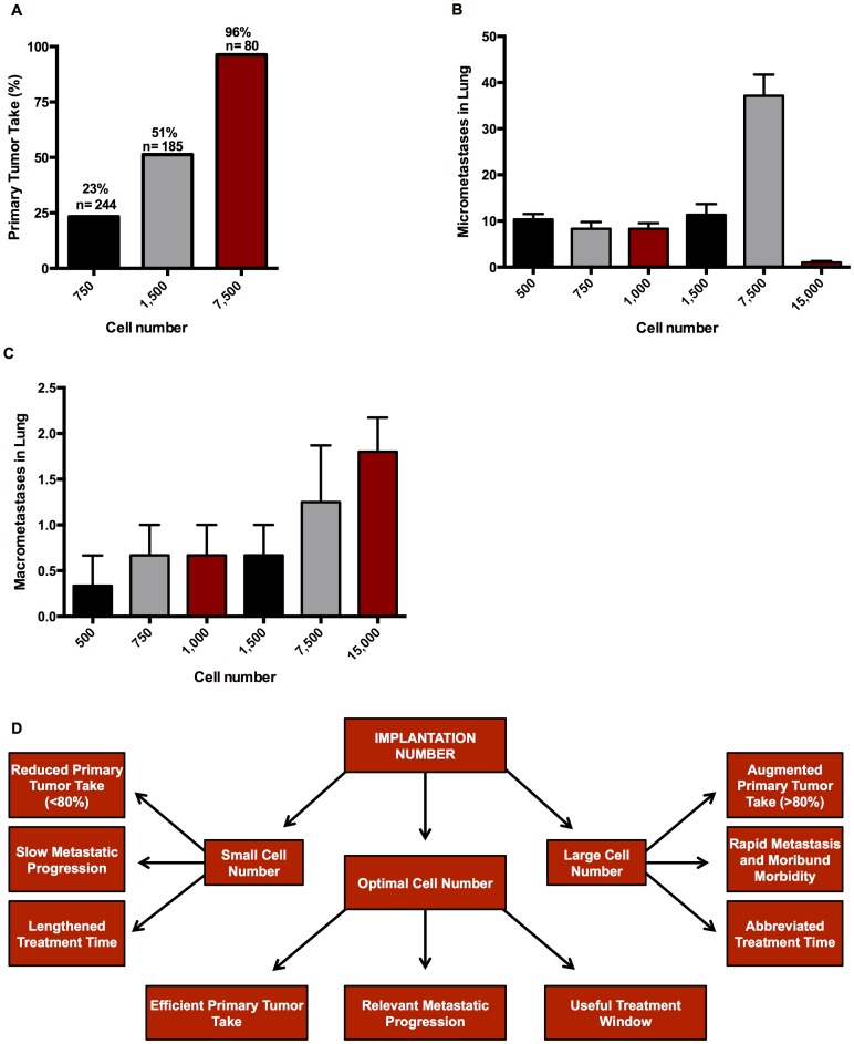
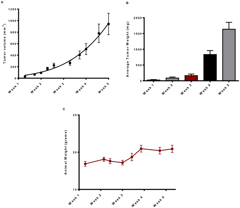
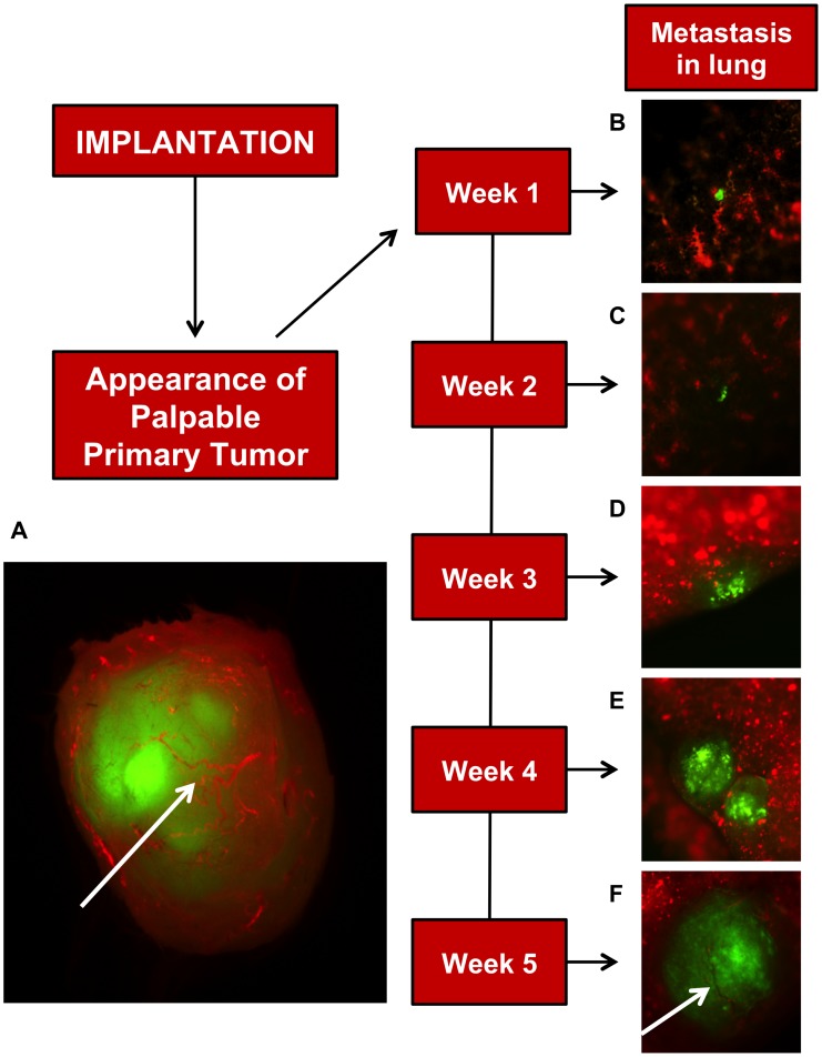
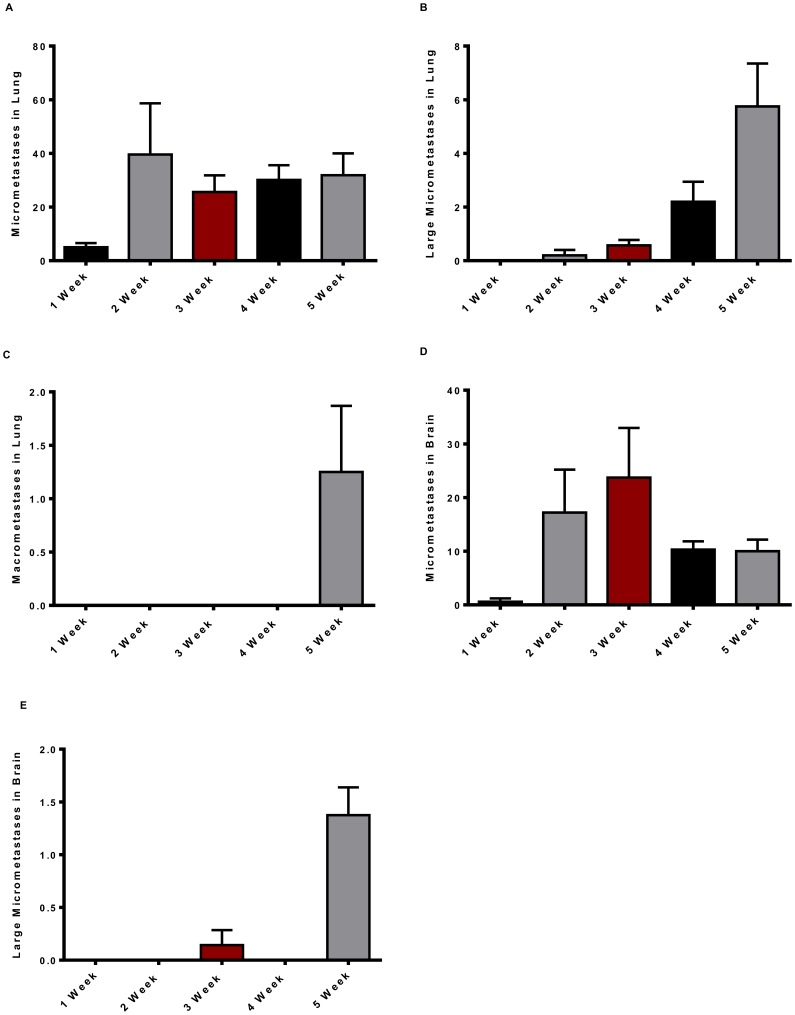
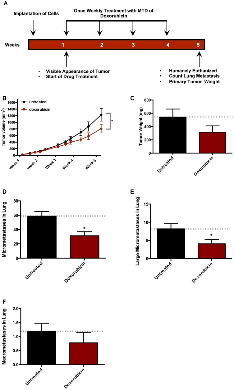
Most cancer patients die with metastatic disease, thus, good models that recapitulate the natural process of metastasis including a dormancy period with micrometastatic cells would be beneficial in developing treatment strategies. Herein we report a model of natural metastasis that balances time to complete experiments with a reasonable dormancy period, which can be used to better study metastatic progression. The basis for the model is a 4T1 triple negative syngeneic breast cancer model without resection of the primary tumor. A cell titration from 500 to 15,000 GFP tagged 4T1 cells implanted into fat pad number four of immune proficient eight week female BALB/cJ mice optimized speed of the model while possessing metastatic processes including dormancy and beginning of reactivation. The frequency of primary tumors was less than 50% in animals implanted with 500-1500 cells. Although implantation with over 10,000 cells resulted in 100% primary tumor development, the tumors and macrometastases formed were highly aggressive, lacked dormancy, and offered no opportunity for treatment. Implantation of 7,500 cells resulted in >90% tumor take by 10 days; in 30-60 micrometastases in the lung (with many animals also having 2-30 brain micrometastases) two weeks post-implantation, with the first small macrometastases present at five weeks; many animals displaying macrometastases at five weeks and animals becoming moribund by six weeks post-implantation. Using the optimum of 7,500 cells the efficacy of a chemotherapeutic agent for breast cancer, doxorubicin, given at its maximal tolerated dose (MTD; 1 mg/kg weekly) was tested for an effect on metastasis. Doxorubicin treatment significantly reduced primary tumor growth and lung micrometastases but the number of macrometastases at experiment end was not significantly affected. This model should prove useful for development of drugs to target metastasis and to study the biology of metastasis.


Conflict of interest statement

Competing Interests: Michael A. Ihnat and Robert E. Hurst are founders and have stock in DormaTarg, Inc. Lora C. Bailey-Downs, Jessica E. Thorpe, Bryan C. Disch, Paul J. Hauser, and Taleah Farasyn are employed by DormaTarg, Inc. DormaTarg, Inc. is a company developed together with the University of Oklahoma in 2007, and the University of Oklahoma has considerable financial interest in this company. Patents 7,575,926 and 8,101,159 have been filed by DormaTarg, Inc., but are not salient to the material in this manuscript. In addition, Dr. Hurst and Michael A. Ihnat receive financial compensation from a small business innovation research (SBIR) grant with DormaTarg. Further, nothing contained within this manuscript represents intellectual property of DormaTarg, Inc. and the model developed herein will be freely available to the scientific community. There are no further patents, products in development or marketed products to declare. This does not alter the authors’ adherence to all the PLOS ONE policies on sharing data and materials, as detailed online in the guide for authors.

Figures

Figure 1
Figure 1. Summary of optimal cell number determination in the development of the metastatic mouse model.
(A) Chart indicating the benefits of optimizing cell number for primary tumor take and metastatic progression in the 4T1 breast cancer model. (B) Percent tumor take in immune-proficient BALB/cJ mice implanted with 4T1-Luc2GFP cells into the number four mammary fat pad. Data are actual percent tumor take based on number of animals indicated above each bar from no fewer than nine independent experiments. Micrometastasis (C) and macrometastasis (D) in lung six weeks after implantation. Data are mean ± SEM from two independent experiments with n = 5 for experiment one, n = 10 for experiment two.
Figure 2
Figure 2. Growth of 4T1-Luc2GFP primary tumors: Timecourse.
Primary tumor growth was recorded over a five week period from BALB/cJ mice implanted with 7,500 4T1-Luc2GFP cells into the mammary fat pad. (A) Tumor volume (mm3). (B) Tumor weight (mg). (C) Animal weights (g). Tumor volumes were calculated using the ellipsoidal method, volume (mm3) = 0.52×length × width2. All data are mean ± SEM from two independent experiments with 5–8 mice for experiment one and 7–11 mice for experiment two and are fitted to a Gomperzian growth curve by Prism 6.0 software.
Figure 3
Figure 3. Development of lung metastasis in the 4T1 Luc2GFP mouse model: Timecourse.
(A) Fluorescent image of a fully vascularized primary tumor removed five weeks after 7,500 4T1-Luc2GFP cells (green in all images) implanted into BALB/cJ mammary fat pad. Vasculature (red) in primary tumor (A) and five weeks lung metastasis (F) are indicated by arrows and was achieved by retro-orbital injection of tetramethylrhodamine labeled 2×10⁶ MW dextran. At weeks one and two (B and C) micrometastases, which are defined as single-to small clusters of cells, are present in lungs. Large micrometastases lacking blood vessels are present in lungs by weeks three and four (D and E), and by week five (F) macrometastases containing visible blood vessels are present in the lungs.
Figure 4
Figure 4. Development and quantification of lung and brain metastases: Timecourse.
Micrometastases examination in lungs and brain of BALB/cJ mice implanted with 7,500 4T1-Luc2GFP cells into mammary fat pad. Lungs and brain excised, examined for metastases, degree of vascularity, and imaged at weeks two-five. (A) Lung micrometastases. (B) Lung large micrometastases. (C) Lung macrometastases. (D) Brain micrometastases. (E) Brain large micrometastases. All data are mean ± SEM from two independent experiments with 5–8 mice for experiment one and 7–11 mice for experiment two.
Figure 5
Figure 5. Efficacy of traditional chemotherapeutic agent in the 4T1 Luc2GPF mouse model.
BALB/cJ mice implanted with 7,500 4T1-Luc2GFP cells were treated with the maximal tolerated dose (MTD) of doxorubicin, the dose shown not to result in any gross toxicity to the animal but shown to result in a small but insignificant loss in weight over the course of the experiment; or with carrier (5% Pharmasolve and 5% Solutol HS in saline), or with injectable saline as control once a week after visible primary tumor formation, for five weeks. (A) Flowchart indicating timing of cell implantation, doxorubicin treatments, and experimental determinations. (B) Tumors volume (mm3) over time measured by calipers. (C) End weight of tumors (mg), (D) Lung micrometastases. (E) Lung large micrometastases. (F) Lung macrometastases. Data representative of two independent experiments n = 15 mice for first experiment, and n = 10 mice for second experiment. Tumor volumes were calculated using the ellipsoidal method, volume (mm3) = 0.52 × length × width2. Data are mean ± SEM and a two way ANOVA/Tukey post-test was performed (*p<0.05 and **p<0.01).

References

    1. Weigelt B, Peterse JL, van’t Veer LJ (2005) Breast cancer metastasis: markers and models. Nat Rev Cancer 5: 591–602 10.1038/nrc1670 - DOI - PubMed
    1. Nkrumah FK, Perkins IV (1976) Relapse in Burkitt’s lymphoma. Int J Cancer 17: 455–460. - PubMed
    1. Yuhas JM, Toya RE, Wagner E (1975) Specific and nonspecific stimulation of resistance to the growth and metastasis of the line 1 lung carcinoma. Cancer Res 35: 242–244. - PubMed
    1. Clayton F, Hopkins CL (1993) Pathologic correlates of prognosis in lymph node-positive breast carcinomas. Cancer 71: 1780–1790. - PubMed
    1. Naumov GN, Townson JL, MacDonald IC, Wilson SM, Bramwell VHC, et al. (2003) Ineffectiveness of doxorubicin treatment on solitary dormant mammary carcinoma cells or late-developing metastases. Breast Cancer Res Treat 82: 199–206 10.1023/B:BREA.0000004377.12288.3c - DOI - PubMed

Publication types

MeSH terms