. 2014 Aug;63(8):847-57.
doi: 10.1007/s00262-014-1561-8. Epub 2014 May 31.

Combination of an agonistic anti-CD40 monoclonal antibody and the COX-2 inhibitor celecoxib induces anti-glioma effects by promotion of type-1 immunity in myeloid cells and T-cells

Affiliations

Combination of an agonistic anti-CD40 monoclonal antibody and the COX-2 inhibitor celecoxib induces anti-glioma effects by promotion of type-1 immunity in myeloid cells and T-cells

Akemi Kosaka et al. Cancer Immunol Immunother. 2014 Aug.

Abstract

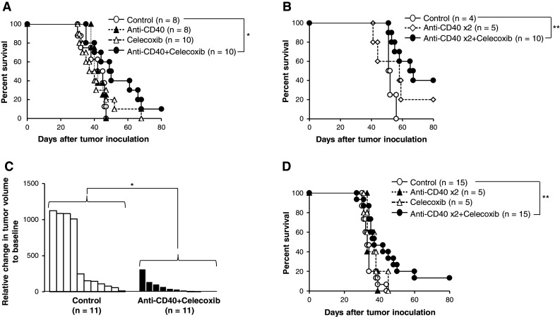
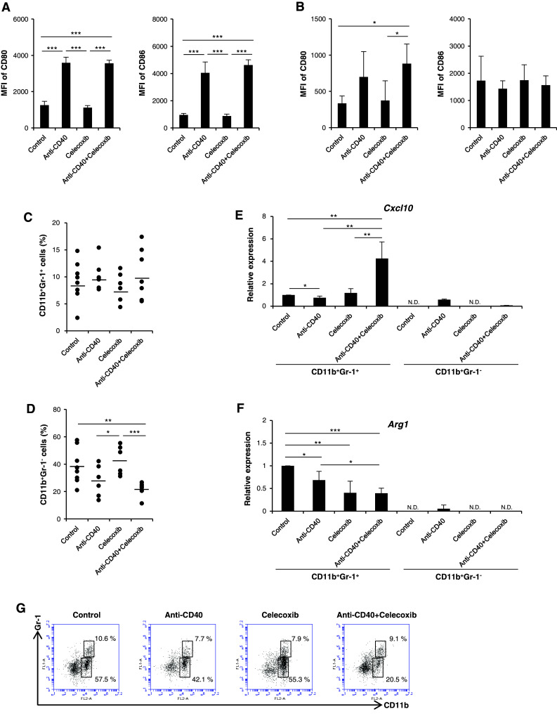
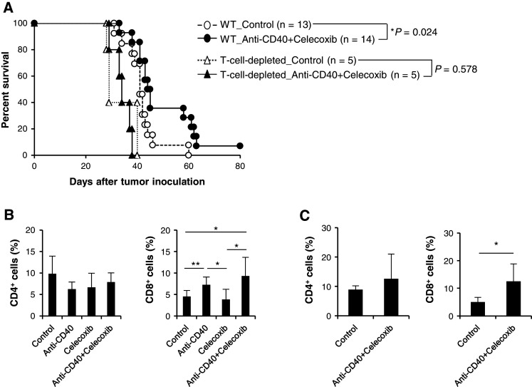
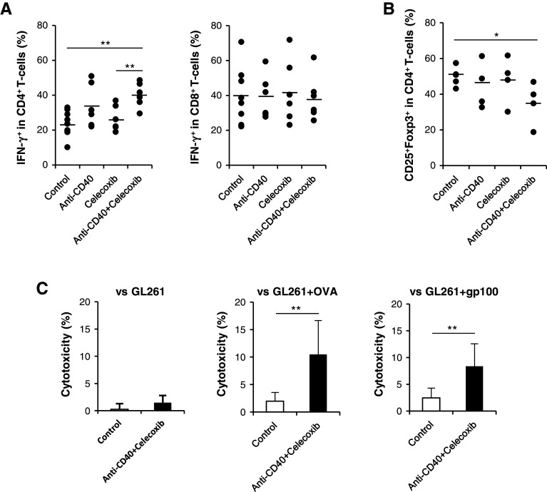
Malignant gliomas are heavily infiltrated by immature myeloid cells that mediate immunosuppression. Agonistic CD40 monoclonal antibody (mAb) has been shown to activate myeloid cells and promote antitumor immunity. Our previous study has also demonstrated blockade of cyclooxygenase-2 (COX-2) reduces immunosuppressive myeloid cells, thereby suppressing glioma development in mice. We therefore hypothesized that a combinatory strategy to modulate myeloid cells via two distinct pathways, i.e., CD40/CD40L stimulation and COX-2 blockade, would enhance anti-glioma immunity. We used three different mouse glioma models to evaluate therapeutic effects and underlying mechanisms of a combination regimen with an agonist CD40 mAb and the COX-2 inhibitor celecoxib. Treatment of glioma-bearing mice with the combination therapy significantly prolonged survival compared with either anti-CD40 mAb or celecoxib alone. The combination regimen promoted maturation of CD11b(+) cells in both spleen and brain, and enhanced Cxcl10 while suppressing Arg1 in CD11b(+)Gr-1(+) cells in the brain. Anti-glioma activity of the combination regimen was T-cell dependent because depletion of CD4(+) and CD8(+) cells in vivo abrogated the anti-glioma effects. Furthermore, the combination therapy significantly increased the frequency of CD8(+) T-cells, enhanced IFN-γ-production and reduced CD4(+)CD25(+)Foxp3(+) T regulatory cells in the brain, and induced tumor-antigen-specific T-cell responses in lymph nodes. Our findings suggest that the combination therapy of anti-CD40 mAb with celecoxib enhances anti-glioma activities via promotion of type-1 immunity both in myeloid cells and T-cells.


Conflict of interest statement

The authors have no financial conflict of interest.

Figures

Fig. 1
Fig. 1
Agonistic anti-CD40 mAb in combination with celecoxib significantly prolonged survival. C57BL/6 mice bearing Quad-GL261 glioma received anti-CD40 mAb i.p. on a day 13 or b days 13 and 23, and/or celecoxib via diet through days 13–33. Kaplan–Meier plot illustrates the survival (*P < 0.05, **P < 0.01, Log-rank test). c C57BL/6 mice bearing established de novo gliomas received the combination therapy or mock treatment. Relative change in tumor volume from day -T1 (baseline; on the day before the treatment started) to day T15 is shown for each mouse as a waterfall plot (n = 11 in each group; *P < 0.05, t test). d C57BL/6 mice bearing SB28 glioma received anti-CD40 mAb i.p. on days 13 and 23 and/or celecoxib via diet through day 13–33. Kaplan–Meier plot illustrates the survival (**P < 0.01, Log-rank test)
Fig. 2
Fig. 2
The combination therapy promoted maturation and M1-like polarization of CD11b+ cells. C57BL/6 mice bearing Quad-GL261 glioma received the combination therapy, monotherapy with anti-CD40 mAb or celecoxib, or mock therapy on day 13 following tumor inoculation. a Spleens and b BILs were harvested on day 15 from individual mice. Expression levels of CD80 and CD86 were evaluated by flow cytometry (n = 5 for control, anti-CD40 and celecoxib groups; n = 4 for the anti-CD40+ celecoxib group; *P < 0.05, ***P < 0.001, t test). c, d Frequencies of CD11b+Gr-1+ and CD11b+Gr-1 cells in the brain were evaluated by flow cytometry on day 5 after therapy (n = 8 for control, n = 6 for monotherapy with anti-CD40 or celecoxib, in each group; n = 7 for anti-CD40+ celecoxib; *P < 0.05, **P < 0.01, ***P < 0.001, t test). e, f BILs were harvested on day 5 posttreatment. CD11b+Gr-1+ and CD11b+Gr-1 BILs were purified from pooled BILs (n = 3–4 in each group) by cell sorting, and mRNA expression levels for Cxcl10 and Arg1 were determined by real-time PCR. Bars and error bars indicate the mean and SD, respectively, from two independent experiments (N.D. not detected; *P < 0.05, **P < 0.01, ***P < 0.001, t test). g Representative flow data of BILs on CD11b+Gr-1+ and CD11b+Gr-1 cells on day 5 posttreatment
Fig. 3
Fig. 3
T-cells were responsible in the observed anti-glioma activity of the combination regimen. a Wild-type (WT) or CD8-deficient mice depleted of CD4+ cells (T-cell-depleted) received intracranial inoculation of Quad-GL261 glioma cells and then received anti-CD40 mAb i.p. on days 13 and 23, and celecoxib via diet through days 13–33. Kaplan–Meier plot illustrates the survival (*P < 0.05, Log-rank test). b C57BL/6 mice bearing Quad-GL261 cells, or c de novo Sleeping Beauty gliomas received anti-CD40 mAb and/or celecoxib. BILs were harvested on day 5 after treatment and analyzed by flow cytometry. Bars and error bars indicate the mean and SD, respectively, from three independent experiments (b n = 8 for control, n = 6 for each of anti-CD40 and celecoxib groups, n = 7 for anti-CD40+ celecoxib, c n = 5 and 6 for control and anti-CD40+ celecoxib, respectively; *P < 0.05, **P < 0.01, t test)
Fig. 4
Fig. 4
The combination of anti-CD40 mAb and celecoxib improved IFN-γ response, reduced regulatory T-cells and induced antigen-specific cytotoxic activities. C57BL/6 mice bearing day 13 Quad-GL261 glioma received the combination, monotherapy with anti-CD40 mAb or celecoxib, or mock treatment. a IFN-γ production and b Foxp3 expression in BILs were determined by flow cytometry on day 18 (a n = 8 for control, n = 6 for each of anti-CD40 and the combination groups; b n = 5 for control, n = 4 for each of anti-CD40 and the combination groups; *P < 0.05, **P < 0.01, t test). c Inguinal lymph node cells were harvested on day 21, and specific cytotoxicity was evaluated against unpulsed GL261 or GL261 cells loaded with OVA257–264 or gp10025–33 peptide by a 6-h 51Cr-release assay at an E:T ratio of 50:1. Data are presented as mean ± SD. (n = 6 per group; **P < 0.01, t test)

Similar articles

Cited by

References

    1. Ostrom QT, Gittleman H, Farah P, Ondracek A, Chen Y, Wolinsky Y, Stroup NE, Kruchko C, Barnholtz-Sloan JS (2013) CBTRUS statistical report: primary brain and central nervous system tumors diagnosed in the United States in 2006-2010. Neuro Oncol 15:ii1–ii56 - PMC - PubMed
    1. Jovčevska I, Kočevar N, Komel R. Glioma and glioblastoma—how much do we (not) know? Mol Clin Oncol. 2013;1:935–941. - PMC - PubMed
    1. Allavena P, Mantovani A. Immunology in the clinic review series; focus on cancer: tumour-associated macrophages: undisputed stars of the inflammatory tumour microenvironment. Clin Exp Immunol. 2012;167:195–205. doi: 10.1111/j.1365-2249.2011.04515.x. - DOI - PMC - PubMed
    1. Schmieder A, Michel J, Schonhaar K, Goerdt S, Schledzewski K. Differentiation and gene expression profile of tumor-associated macrophages. Semin Cancer Biol. 2012;22:289–297. doi: 10.1016/j.semcancer.2012.02.002. - DOI - PubMed
    1. Khaled YS, Ammori BJ, Elkord E. Myeloid-derived suppressor cells in cancer: recent progress and prospects. Immunol Cell Biol. 2013;91:493–502. doi: 10.1038/icb.2013.29. - DOI - PubMed

Publication types

MeSH terms