

Dose response effect of Paracoccidioides brasiliensis in an experimental model of arthritis
- PMID: 24879005
- PMCID: PMC4085864
- DOI: 10.1590/s0036-46652014000300012
Dose response effect of Paracoccidioides brasiliensis in an experimental model of arthritis
Abstract
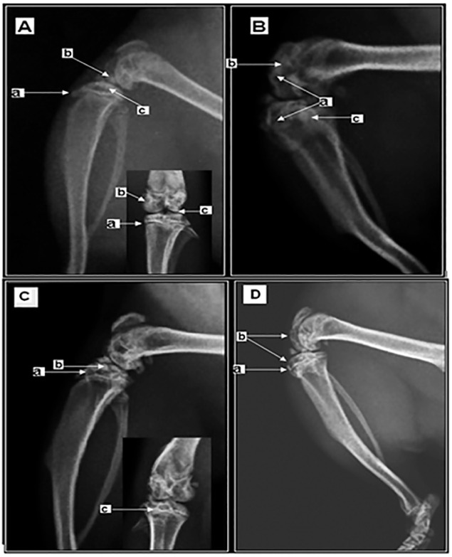
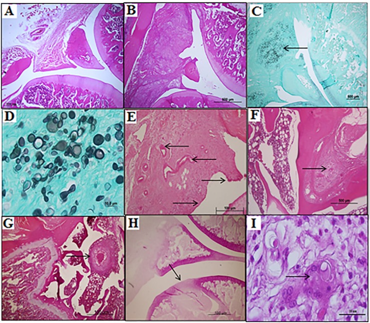
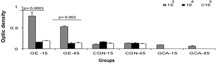
Paracoccidioidomycosis (PCM) is caused by the dimorphic fungus Paracoccidioides brasiliensis (Pb) and corresponds to prevalent systemic mycosis in Latin America. The aim of the present work was to evaluate the dose response effect of the fungal yeast phase for the standardization of an experimental model of septic arthritis. The experiments were performed with groups of 14 rats that received doses of 103, 104 or 105 P. brasiliensis (Pb18) cells. The fungi were injected in 50 µL of phosphate-buffered saline (PBS) directly into the knee joints of the animals. The following parameters were analyzed in this work: the formation of swelling in knees infused with yeast cells and the radiological and anatomopathological alterations, besides antibody titer by ELISA. After 15 days of infection, signs of inflammation were evident. At 45 days, some features of damage and necrosis were observed in the articular cartilage. The systemic dissemination of the fungus was observed in 11% of the inoculated animals, and it was concluded that the experimental model is able to mimic articular PCM in humans and that the dose of 105 yeast cells can be used as standard in this model.
A paracoccidioidomicose (PCM) é causada pelo fungo dimórfico Paracoccidioides brasiliensis (Pb) e corresponde à micose sistêmica de maior prevalência na América Latina. O objetivo do presente trabalho foi avaliar a dose resposta de leveduras do fungo para padronização do modelo experimental de artrite séptica. Os experimentos foram realizados com grupos de 14 ratos que receberam doses de 103, 104 ou 105 células de P. brasiliensis (Pb18). Os fungos foram injetados em 50 µL de solução salina em tampão fosfatado (PBS) diretamente na articulação do joelho dos animais. Os seguintes parâmetros foram analisados neste trabalho: a formação de edema nos joelhos infundidos com as células das leveduras e alterações radiológicas, anatopalógicas além de titulação de anticorpos por Elisa. Após 15 dias de infecção, os sinais de inflamação foram evidentes. Aos 45 dias, algumas características de dano e necrose foram observadas na cartilagem articular. A disseminação sistêmica do fungo foi observada em 11% dos animais inoculados, concluiu-se que o modelo experimental é capaz de mimetizar a PCM articular em humanos e que a dose de 105 leveduras representa a dose padrão para o desenvolvimento do modelo.
Figures
Similar articles
-
Natural history of experimental arthritis induced by Paracoccidioides brasiliensis in wistar rats.Rev Soc Bras Med Trop. 2019 Jan 14;52:e20180043. doi: 10.1590/0037-8682-0043-2018. Rev Soc Bras Med Trop. 2019. PMID: 30652784
-
Experimental model of arthritis induced by Paracoccidioides brasiliensis in rats.Mycopathologia. 2012 Sep;174(3):187-91. doi: 10.1007/s11046-012-9537-8. Epub 2012 Mar 30. Mycopathologia. 2012. PMID: 22460985
-
Distinct patterns of yeast cell morphology and host responses induced by representative strains of Paracoccidioides brasiliensis (Pb18) and Paracoccidioides lutzii (Pb01).Med Mycol. 2016 Feb;54(2):177-88. doi: 10.1093/mmy/myv072. Epub 2015 Sep 17. Med Mycol. 2016. PMID: 26384386
-
[The Research Encouragement Award. Effects of sex hormones on sexual difference of experimental paracoccidioidomycosis].Nihon Ishinkin Gakkai Zasshi. 1999;40(1):1-8. doi: 10.3314/jjmm.40.1. Nihon Ishinkin Gakkai Zasshi. 1999. PMID: 9929575 Review. Japanese.
-
Functional genome of the human pathogenic fungus Paracoccidioides brasiliensis.FEMS Immunol Med Microbiol. 2005 Sep 1;45(3):369-81. doi: 10.1016/j.femsim.2005.05.013. FEMS Immunol Med Microbiol. 2005. PMID: 16061364 Review.
Cited by
-
Osteoarticular Mycoses.Clin Microbiol Rev. 2022 Dec 21;35(4):e0008619. doi: 10.1128/cmr.00086-19. Epub 2022 Nov 30. Clin Microbiol Rev. 2022. PMID: 36448782 Free PMC article. Review.
References
-
- Amanai T, Nakamura Y, Aoki S, Mataga I. Micro-CT analysis of experimental Candida osteoarthritis in rats. Mycopathologia. 2008;166:133–41. - PubMed
-
- Baida H, Biselli PJ, Juvenale M, Del Negro GM, Mendes-Giannini MJ, Duarte AJ, et al. Differential antibody isotype expression to the major Paracoccidioides brasiliensis antigen in juvenile and adult form paracoccidioidomycosis. Microbes Infect. 1999;1:273–8. - PubMed
-
- Baransky MC, Silva AF, Rodrigues D. Lesões ósseas e osteoarticulares. In: Del Negro G, Lacaz CS, Fiorillo AM, editors. Paracoccidioidomicose: blastomicose sul-americana. São Paulo: Sarvier-Edusp; 1982. pp. 211–9.
-
- Benard G, Mendes-Giannini MJ, Juvenale M, Miranda ET, Duarte AJ. Immunosuppression in paracoccidioidomycosis: T cell hyporesponsiveness to two Paracoccidioides brasiliensis glycoproteins that elicit strong humoral immune response. J Infect Dis. 1997;175:1263–7. - PubMed
Publication types
MeSH terms
LinkOut - more resources
Full Text Sources
Other Literature Sources
Medical
