. 2014 Dec;21(6):589-95.
doi: 10.1007/s10140-014-1243-z. Epub 2014 May 31.

The accuracy of pre-appendectomy computed tomography with histopathological correlation: a clinical audit, case discussion and evaluation of the literature

Affiliations

The accuracy of pre-appendectomy computed tomography with histopathological correlation: a clinical audit, case discussion and evaluation of the literature

George Benjamin Collins et al. Emerg Radiol. 2014 Dec.

Abstract

The increasing use of computed tomography (CT) in acute appendicitis makes recognising the radiological hallmarks of the condition and its mimics vital. The differential diagnosis includes both appendiceal and nonappendiceal pathologies. The correlation between pre-appendectomy CT and post-appendectomy histopathology was audited retrospectively. Cases of clinico-histopathological discrepancy underwent blind peer-review, and possible improvements were discussed in the context of the medical literature. A grade for discrepancy was given based on the RADPEER scoring system, and interesting or discrepant cases were examined more closely to identify targets for education. Of the 199 procedures, 4 appendectomies were negative (histologically normal), 182 were positive (primary appendicitis) and 13 were incidental (another primary process caused inflammation). The positive predictive value for pre-appendectomy CT was 91.5 %, and the negative appendectomy rate was 2 %. There were many secondary pathologies, including neoplasia, tuberculosis and endometriosis. Although no CT reports missed a diagnosis that should be made "almost all of the time" and in 96 % of cases, the second, blinded radiologist agreed with the initial assessment, in 3 cases, a missed diagnosis altered clinical management; 2 were "understandable" misses but 1 was not. In five cases, a discrepancy was "understandable" but clinically insignificant. Overall, in comparison to the medical literature, the degree of clinico-histopathological correlation was good. Although identifying areas for improvement was challenging, after a pictorial review of four cases and a discussion of the medical literature, we present our audit results and some valuable learning points for use in the CT assessment of suspected acute appendicitis.


Figures
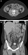
Fig. 1
Fig. 1
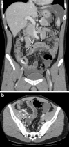
A 16-year-old male with mesenteric lymphadenitis mimicking acute appendicitis (case 1). Contrast-enhanced axial (a) and coronal (b) images demonstrate prominent enlarged ileocolic lymph notes (white arrows). The appendix could not be identified due to paucity of intra-abdominal fat.
Fig. 2
Fig. 2
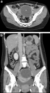
a A 32-year-old male with enteritis masquerading as acute appendicitis (case 2; see b). Contrast-enhanced coronal images demonstrate a fluid-filled small bowel loop with mural thickening and enhancement in keeping with enteritis (white arrows). Ascites is noted within the perisplenic region and pelvis. b A 32-year-old male with enteritis (case 2; see b). Contrast-enhanced axial CT image demonstrates a small air locule (white arrow) at the tip of a blind-ending tubular structure (open white arrows) within the right iliac fossa with a slightly thickened and enhancing wall, suggesting possible perforation of an acutely inflamed appendix. Intraluminal dense material was in keeping with an appendicolith (black arrow)
Fig. 3
Fig. 3
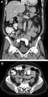
a A 35-year-old female with oophoritis in early pelvic inflammatory disease mimicking an acute appendicitis (case 3; see a). The contrast-enhanced axial CT scan image reveals incidental bilateral ovarian cysts, larger on the left (white asterisks). There are small pockets of free fluid and inflammatory fat stranding surrounding the ovarian cysts (white arrows). b A 35-year-old female with oophoritis in early pelvic inflammatory disease (case 3; see a). In this contrast-enhanced coronal CT image, there is a suggestion of mild focal mural thickening and enhancement of the retrocaecal appendix (white arrows), associated with a small amount of periappendiceal fluid
Fig. 4
Fig. 4
A 64-year-old male with moderately differentiated adenocarcinoma of the appendix (case 4). The coronal (a) and axial (b) contrast-enhanced CT scan images reveal a dilated, fluid-filled appendix with mural thickening and enhancement (white arrows). There is also evidence of periappendiceal inflammatory fat stranding (white asterisk) which is best appreciated in the axial image

Similar articles

Cited by

References

    1. Dude JB, Lynch ML, Bhatt S, Dogra VS. Computed tomography mimics of acute appendicitis: predictors of appendiceal disease confirmed at pathology. J Clin Imaging Sci. 2012;2(73):1–8. - PMC - PubMed
    1. Otero HJ, Ondategui-Parra S, Erturk SM, Ochoa RE, Gonzalez-Beicos A, Ros PR. Imaging utilization in the management of appendicitis and its impact on hospital charges. Emerg Radiol. 2008;15(1):23–28. doi: 10.1007/s10140-007-0678-x. - DOI - PubMed
    1. Heller MT, Hattoum A. Imaging of acute right lower quadrant abdominal pain: differential diagnoses beyond appendicitis. Emerg Radiol. 2012;19(1):61–73. doi: 10.1007/s10140-011-0997-9. - DOI - PubMed
    1. Jackson VP, Cushing T, Abujudeh HH, Borgstede JP, Chin KW, Grimes CK, Larson DB, Larson PA, Pyatt RS, Thorwarth WT. RADPEER™ scoring white paper. J Am Coll Radiol. 2009;6(1):21–25. doi: 10.1016/j.jacr.2008.06.011. - DOI - PubMed
    1. Whitley S, Sookur P, McLean A, Power N. The appendix on CT. Clin Radiol. 2009;64(2):190–199. doi: 10.1016/j.crad.2008.06.015. - DOI - PubMed