. 2014 Jun;10(6):957-71.
doi: 10.4161/auto.28363.

Identification of a novel MTOR activator and discovery of a competing endogenous RNA regulating autophagy in vascular endothelial cells

Affiliations

Identification of a novel MTOR activator and discovery of a competing endogenous RNA regulating autophagy in vascular endothelial cells

Di Ge et al. Autophagy. 2014 Jun.

Abstract

MTOR, a central regulator of autophagy, is involved in cancer and cardiovascular and neurological diseases. Modulating the MTOR signaling balance could be of great significance for numerous diseases. No chemical activators of MTOR have been found, and the urgent challenge is to find novel MTOR downstream components. In previous studies, we found a chemical small molecule, 3-benzyl-5-((2-nitrophenoxy) methyl)-dihydrofuran-2(3H)-one (3BDO), that inhibited autophagy in human umbilical vein endothelial cells (HUVECs) and neuronal cells. Here, we found that 3BDO activated MTOR by targeting FKBP1A (FK506-binding protein 1A, 12 kDa). We next used 3BDO to detect novel factors downstream of the MTOR signaling pathway. Activation of MTOR by 3BDO increased the phosphorylation of TIA1 (TIA1 cytotoxic granule-associated RNA binding protein/T-cell-restricted intracellular antigen-1). Finally, we used gene microarray, RNA interference, RNA-ChIP assay, bioinformatics, luciferase reporter assay, and other assays and found that 3BDO greatly decreased the level of a long noncoding RNA (lncRNA) derived from the 3' untranslated region (3'UTR) of TGFB2, known as FLJ11812. TIA1 was responsible for processing FLJ11812. Further experiments results showed that FLJ11812 could bind with MIR4459 targeting ATG13 (autophagy-related 13), and ATG13 protein level was decreased along with 3BDO-decreased FLJ11812 level. Here, we provide a new activator of MTOR, and our findings highlight the role of the lncRNA in autophagy.

Keywords: ATG13; MIR4459; MTOR activator; TIA1; autophagy; lncRNA.


Figures

None
Figure 1. 3BDO failed to activate the MTOR signaling in FKBP1A protein-overexpressed HUVECs. (A) In silico docking of 3BDO into the hydrophobic pocket of FKBP1A and surface view of docked 3BDO-FKBP1A molecule. Hydrogen-bond network of the docked 3BDO-FKBP1A molecule. (B) Western blot analysis of EIF4EBP1, phosphorylated EIF4EBP1 (p-EIF4EBP1, Thr37/46), RPS6KB1, and phosphorylated RPS6KB1 (p-RPS6KB1, Thr389) in HUVECs treated with or without 3BDO (60 μM) for 24 h after overexpression of FKBP1A. (C) Densitometry results of ratio of p-RPS6KB1 to total RPS6KB1 and p-EIF4EBP1 to total EIF4EBP1. pCMV6 phosphorylation was set to 1. *P < 0.05, **P < 0.01, #P > 0.05, n = 3.
None
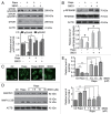
Figure 2. 3BDO inhibited the role of rapamycin in the MTOR pathway and autophagy. (A) Western blot analysis of the phosphorylation of MTOR (Ser2448 and Ser2481) in HUVECs treated with 3BDO (60 μM), rapamycin (10 μM) or both for 6 h. Densitometry results are the ratio of p-MTOR (Ser2448 and Ser2481) to MTOR, with data for the nontreated group set to 1. (B) Western blot analysis of substrates of MTOR signaling in HUVECs, pretreated with or without 3BDO (60 μM) for 30 min, then stimulated with rapamycin (10 μM) for 6 h. (C) Images of MAP1LC3B dots in HUVEC. HUVECs were pretreated with or without 3BDO (60 μM) for 30 min, then stimulated with rapamycin (10 μM) for 6 h. Scale bar: 16 μm. (D) Western blot analysis of MAP1LC3B and SQSTM1 protein levels in HUVEC. HUVECs were pretreated with or without 3BDO (7.5, 15 or 30 μM) for 30 min, then stimulated with rapamycin (10 μM) for 6 h. (E) Densitometry results of ratio of SQSTM1 to ACTB. Ctr data were set to 1. Densitometry results of ratio of MAP1LC3B-II to ACTB. Ctr data were set to 1. *P < 0.05, **P < 0.01, n = 3.
None
Figure 3. 3BDO promoted phosphorylation of TIA1 in HUVECs. HUVECs were treated with rapamycin (10 μΜ), 3BDO (60 μΜ) or both for 6 h; immunoprecipitation was obtained with TIA1 antibody. Western blot analysis of phosphorylation of Ser residues of TIA1. Densitometry results of the ratio of p-Ser to TIA1 with data for the nontreated group set to 1. *P < 0.05, n = 3.
None
Figure 4.FLJ11812 found on microarray analysis and was an lncRNA derived from the 3′UTR of TGFB2. (A) Differentially expressed transcripts induced by 60 μM 3BDO for 24 h. The scatter plot depicts genes with > 2-fold change in expression: upregulation (red) and downregulation (green). The arrow shows FLJ11812 with the most downregulated expression. (B) UCSC Genome Bioinformatics Browser data for FLJ11812 transcript on chromosome band 1p41. And gene view of TGFB2 indicating coding sequence (CDS, blue), 3′UTR and location of FLJ11812 (red) according to gene annotation of TGFB2. (C) (Top panel) Positions of probes for TGFB2 and FLJ11812. (Bottom panel) northern blot analysis of FLJ11812 and TGFB2 with specific probes against FLJ11812 or TGFB2. (D) Quantitative PCR (qPCR) analysis of mRNA expression of FLJ11812 and its adjacent subregion in the 3′UTR of TGFB2. (Top panel) Gene view of TGFB2 3′UTR indicating CDS (black), 3′UTR (green) and location of FLJ11812 (yellow) according to gene annotation of TGFB2. (Bottom panel) qPCR analysis of FLJ11812 with a series of corresponding primer pairs. Every span is the subregion of the TGFB2 3′UTR sequence.
None
Figure 5. 3BDO could downregulate FLJ11812 and did not affect TGFB2 expression. (A) Positions of the 2 primers used in qPCR. (B) RT-PCR analysis of the level of FLJ11812 in HUVECs treated with different concentrations of 3BDO for 24 h and with 120 μM 3BDO for various times. (C) qPCR analysis of the mRNA level of TGFB2 in HUVECs treated with concentrations of 3BDO for 24 h and 120 μM 3BDO for various times. (D) Western blot analysis of TGFB2 protein level and quantification. (E and F) qPCR analysis of levels of FLJ11812 and TGFB2 in HUVECs treated with scrambled siRNA or FLJ11812 siRNA for 36 h. *P < 0.05. **P < 0.01, n = 3.
None
Figure 6. TIA1 regulated the processing of FLJ11812. (A) Western blot analysis of siRNA knockdown of TIA1. (B and C) qPCR analysis of the mRNA levels of FLJ11812 and TGFB2 in HUVECs with siRNA knockdown of TIA1 for 36 h. **P < 0.01, #P > 0.05, n = 3. (D) HUVECs were treated with DMSO or 120 μM 3BDO for 10 h, then total RNA-protein complexes purification were performed using these cells and underwent ChIP assay. ChIP assays with TIA1 antibody against TIA1 and the fragment that contains part of the FLJ11812 sequence amplified by RT-PCR with an adjusted PCR cycle with specific primer pairs. (E) Positions of the 2 primers used in RNA-ChIP experiments. Position 1 is the TIA1 binding sequence, and position 2 is the negative control sequence.
None
Figure 7.FLJ11812 induced HUVEC autophagy. (A) Western blot analysis of MAP1LC3B protein level in HUVECs transfected with pCMV6 or pCMV6-FLJ11812 for 48 h. (B and C) Immunostaining of MAP1LC3B in HUVECs transfected with pCMV6 or pCMV6-FLJ11812 (0.2 μg/ml) for 48 h, and the percentage of cells containing MAP1LC3B puncta (> 5). Scale bar: 16 μm. Western blot analysis of protein level of (D) MAP1LC3B in HUVECs transfected with pCMV6 or pCMV6-FLJ11812 (0.2 μg/ml) for 42 h, then treated with or without bafilomycin for 6 h; (E) MAP1LC3B in HUVECs transfected with pCMV6 or pCMV6-FLJ11812 (0.2 μg/ml) for 24 h, then treated with or without 3BDO for 24 h; and (F) TGFB2 in HUVECs transfected with pCMV6 and pCMV6-FLJ11812 for 48 h. *P < 0.05, **P < 0.01, #P > 0.05, n = 3.
None
Figure 8.FLJ11812 is a natural decoy for MIR4459. (A) Potential sites in Luc-FLJ11812 targeted by MIR4459. FLJ11812 fragments and mutant derivatives devoid of MIR4459 binding-site fragments were cloned into the luciferase reporter vector, thus resulting in 2 constructs, Luc-FLJ11812-WT and Luc-FLJ11812-Δ4459 constructs. (B) Luc-FLJ11812-WT plasmid was cotransfected into HEK293 cells with different concentrations of MIR4459 or scrambled miRNA for 24 h and luciferase activity was measured. (C) Luc-FLJ11812-WT and Luc-FLJ11812-Δ4459 plasmids were transfected into HEK293 cells with scrambled miRNA or MIR4459 for 24 h and luciferase activity was measured. (D and E) Potential sites in the 3′UTR of ATG13 targeted by MIR4459. The 3′UTR of ATG13 fragment was cloned into the luciferase reporter vector, resulting in the Luc-ATG13-WT construct. This construct was cotransfected into HEK293 cells with different concentrations of MIR4459 or a control miRNA for 24 h, then luciferase activity was detected. *P < 0.05, **P < 0.01, #P > 0.05, n = 3.
None
Figure 9.FLJ11812 decreased the level of MIR4459, and MIR4459 suppressed ATG13 protein level. (A) qPCR analysis of MIR4459 level in HUVECs treated with or without 3BDO for 24 h. (B) qPCR analysis of MIR4459 level in HUVECs transfected with pCMV6, pCMV6-FLJ11812 or MIR4459-insensitive mutant of FLJ11812 (Mut-FLJ11812) for 48 h. Western blot analysis of protein level of (C) ATG13 in MIR4459-transduced HUVECs; (D) ATG13 in HUVECs transfected with pCMV6 or pCMV6-FLJ11812 for 48 h (densitometry results are the ratio of ATG13 to ACTB or GAPDH); and (E) p-ATG13 (Ser318) and ATG13 in HUVEC stimulated with rapamycin (10 μM) for 6 h after pretreatment with or without 3BDO for 30 min. *P < 0.05, **P < 0.01, n = 3.
None
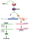
Figure 10. Conceptual schematic of 3BDO and FLJ11812 mechanism of action. 3BDO can occupy the rapamycin-binding site, blocks the interaction of rapamycin and FKBP1A, and activates the MTOR signaling pathway and subsequent TIA1 phosphorylation. TIA1 phosphorylation and the reduced interaction between the 3′ UTR of TGFB2 and TIA1 by 3BDO might affect the production of an lncRNA, FLJ11812. FLJ11812, with decoy activity for MIR4459 and by sequestering MIR4459, regulates the level of MIR4459 target, ATG13, and autophagy.

Similar articles

Cited by

References

    1. Mizumura K, Cloonan SM, Haspel JA, Choi AM. The emerging importance of autophagy in pulmonary diseases. Chest. 2012;142:1289–99. doi: 10.1378/chest.12-0809. - DOI - PMC - PubMed
    1. Hay N, Sonenberg N. Upstream and downstream of mTOR. Genes Dev. 2004;18:1926–45. doi: 10.1101/gad.1212704. - DOI - PubMed
    1. Tsang CK, Qi H, Liu LF, Zheng XF. Targeting mammalian target of rapamycin (mTOR) for health and diseases. Drug Discov Today. 2007;12:112–24. doi: 10.1016/j.drudis.2006.12.008. - DOI - PubMed
    1. Zaytseva YY, Valentino JD, Gulhati P, Evers BM. mTOR inhibitors in cancer therapy. Cancer Lett. 2012;319:1–7. doi: 10.1016/j.canlet.2012.01.005. - DOI - PubMed
    1. Weichhart T. Mammalian target of rapamycin: a signaling kinase for every aspect of cellular life. Methods Mol Biol. 2012;821:1–14. doi: 10.1007/978-1-61779-430-8_1. - DOI - PubMed

Publication types

MeSH terms

Substances