. 2014 Jul 15;98(1):29-38.
doi: 10.1097/TP.0000000000000170.

Heat shock protein-27 delays acute rejection after cardiac transplantation: an experimental model

Affiliations

Heat shock protein-27 delays acute rejection after cardiac transplantation: an experimental model

Borggia Seemampillai et al. Transplantation. .

Abstract

Background: Rejection is the major obstacle to survival after cardiac transplantation. We investigated whether overexpression of heat shock protein (Hsp)-27 in mouse hearts protects against acute rejection and the mechanisms of such protection.

Methods: Hearts from B10.A mice overexpressing human Hsp-27 (Hsp-27tg), or Hsp-27-negative hearts from littermate controls (LCs) were transplanted into allogeneic C57BL/6 mice. The immune response to B10.A hearts was investigated using quantitative polymerase chain reaction for CD3+, CD4+, CD8+ T cells, and CD14+ monocytes and cytokines (interferon-γ, interleukin [IL]-2, tumor necrosis factor-α, IL-1β, IL-4, IL-5, IL-10, transforming growth factor-β) in allografts at days 2, 5, and 12 after transplantation. The effect of Hsp-27 on ischemia-induced caspase activation and immune activation was investigated.

Results: Survival of Hsp-27tg hearts (35±10.37 days, n=10) was significantly prolonged compared with LCs (13.6±3.06 days, n=10, P=0.0004). Hsp-27tg hearts expressed significantly more messenger RNA (mRNA) markers of CD14+ monocytes at day 2 and less mRNA markers of CD3+ and CD8+T cells at day 5 compared with LCs. There was more IL-4 mRNA in Hsp-27tg hearts at day 2 and less interferon-γ mRNA at day 5 compared with LCs. Heat shock protein-27tg hearts subjected to ischemia or to 24 hr ischemia-reperfusion injury demonstrated significantly less apoptosis and activation of caspases 3, 9, and 1 than LCs. T cells removed from C57BL/6 recipients of Hsp-27tg hearts produced a vigorous memory response to B10.A antigens, suggesting immune activation was not inhibited by Hsp-27.

Conclusion: Heat shock protein-27 delays allograft rejection, by inhibiting tissue damage, through probably an antiapoptotic pathway. It may also promote an anti-inflammatory subset of monocytes.


Conflict of interest statement

The authors declare no conflicts of interest.

Figures

FIGURE 1
FIGURE 1
Distribution of HA-tagged Hsp-27 in transgenic mice and prolonged survival of transgenic allograft. Protein from hearts, lungs, liver, kidney and spleen from LC and Hsp-27tg mice were separated by SDS-PAGE and transferred to nitrocellulose membranes by Western blotting (a). They were probed with antibodies to HA, Hsp-25 or GAPDH followed by HRP-conjugated antibody. In b, frozen sections of hearts from Hsp-27tg and LCs were treated with peroxidase labeled irrelevant antibody (neg control) or antibody to HA, the peroxidase was visualized as described in the Materials and Methods. Hemagglutinin is predominantly expressed within cardiomyocytes of Hsp-27tg mice. In c, sera from LCs (n=8) and Hsp-27tg mice (n=8) were tested for soluble Hsp-27 by ELISA. The concentration of Hsp-27 was determined using the standard curves provided by the manufacture. Results are shown as mean±SEM, P=<0.001. In d, Hearts from Hsp-27tg (n=10) or their LCs (n=10) were heterotopically transplanted in C57BL/6 recipients. Survival of hearts from Hsp-27tg mice were significantly prolonged compared with hearts from LC (P<0.001, by Kaplan-Meier analysis). Syngeneic hearts were still beating at day 50 when all surviving mice were killed. HA, hemagglutinin; SDS-PAGE, sodium dodecyl sulfate-polyacrylamide gel electrophoresis; LCs, littermate controls; Hsp, heat shock protein. ELISA, enzyme-linked immunosorbent assay.
FIGURE 2
FIGURE 2
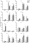
Effect of Hsp-27 on infiltrating cells. Recipients of transgenic and LC allografts were killed at days 2, 5, and 12 after transplantation and donor hearts were assessed for infiltration of CD3+ (A), CD4+ (B), and CD8+ (C) T cells as well as CD14+ (D) cells by RT-PCR. The y-axis shows relative expression of mRNA as a percentage of that present in syngrafts. It can be seen that transgenic allografts have less CD3+ and CD8+ T cells at day 5 compared with LC allografts and more CD14 cells at day 2 (* or #P≤0.05; ** or ##P<0.01 and *** or ###P<0.001, *transgenic vs. LC, #syngeneic vs. LC or syngeneic vs. transgenic, n=3–6). LC, littermate control; mRNA, messenger RNA; RT-PCR, reverse-transcriptase polymerase chain reaction; Hsp, heat shock protein.
FIGURE 3
FIGURE 3
Effect of Hsp-27 on cytokines. Recipients of transgenic and LC allografts were killed at days 2, 5, and 12 after transplantation and donor hearts assessed for presence of IFN-γ (A), IL-2 (B), TNF-α (C), IL-1β (D), IL-4 (E), IL-5 (F), IL-10 (g) and TGF-β (H). The y-axis shows relative expression of mRNA as a percentage of that present in syngrafts. It can be seen that transgenic allografts express less IFN-γ at day 5 than LC allografts and more IL-4 at day 2 (*or #P≤0.05; ** or ##P<0.01 and *** or ###P<0.001, *transgenic vs. LC, #syngeneic vs. LC or syngeneic versus transgenic, n=3–6). IL, interleukin; TNF, tumor necrosis factor; TGF, transforming growth factor; mRNA, messenger RNA; LC, littermate control; IFN, interferon; Hsp, heat shock protein.
FIGURE 4
FIGURE 4
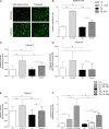
Comparison of apoptotic cells and caspase activity induced by ischemia-reperfusion injury in Hsp-27tg and LC hearts. Hearts from Hsp-27tg (n=3–6) and LC (n=3–6) mice were subjected to 10 min cold ischemia and 40 min warm ischemia ex vivo (A–E) or ischemia-reperfusion injury in vivo for 4 hr or 24 hr (F). The hearts from these mice were subjected to TUNEL assay (a and b) to reveal presence of apoptotic cells or examined for presence of caspases. A, A photomicrograph of cryostat sections of hearts from Hsp-27tg and LC hearts (normoxic and ischemic) showing green fluorescent TUNEL-positive apoptotic cells. B, Quantitation of apoptotic cells (y-axis — no of apoptotic cells) in hearts from nonischemic LC, LC ischemic, nonischemic Hsp-27tg and ischemic Hsp-27tg. Data derived from mean of 50 fields/heart. Heat shock protein-27tg hearts subjected to ischemia had less apoptotic cells than LC hearts subjected to ischemia. (C–E) Activity of caspase 3 (n=6 per group), caspase 9 (n=5 per group), and caspase 1 (n=4 per group), respectively, after ischemia measured as OD460/min/μg. It can be seen that ischemia-induced expression of caspase 3 and caspase 9 is significantly reduced in Hsp-27tg compared with LC hearts (C and D). The x-axis of f (n=3) shows the groups (LC normoxic, LC after 4 or 24 hr ischemia-reperfusion, Hsp-27tg normoxic, Hsp-27tg after 4 or 24 hr ischemia-reperfusion). It can be seen that there was no significant increase in caspase 3 at 4 hr in any group, but at 24 hr, there was significantly more caspase activity in LC hearts compared with normoxic hearts. One-way ANOVA with Bonferroni correction (*P<0.05, **P<0.01, and ***P<0.001). LC, littermate control; TUNEL, terminal deoxynucleotide transferase-mediated 2’-deoxyuridine-5’-triphosphate nick-end labeling; ANOVA, analysis of variance; Hsp, heat shock protein.
FIGURE 5
FIGURE 5
Frequency of alloreactive T cells from mice receiving Hsp-27tg and LC hearts grafts. Splenic T cells obtained from C57BL/6 recipients of syngeneic, LC, or Hsp-27tg allografts were removed at 5 (A) or 12 days (B) after transplantation and mixed with antigen-presenting cell (APC) isolated from LC, Hsp-27tg, third party (FVB) controls or medium alone. The cultures were tested for production of IFN-γ by ELISPOT. The frequency of IFN-γ–producing cells is shown on the y-axis as number of spots per million splenocytes. Splenocytes from recipients of LC or Hsp-27tg allografts demonstrated a vigorous memory response when cultured in the presence of APC from LC or Hsp-27tg mice, but exhibited a poor response to APC from third party controls. There is no evidence that the presence of the Hsp-27tg allograft for 5 or 12 days diminishes the ability of C57BL/6 splenocytes to become primed to B10.A antigens, *P<0.05, **P<0.01, ***P<0.001, n=3–8 per group. LC, littermate control; APC, antigen-presenting cell; IFN, interferon; ELISPOT, enzyme-linked immunosorbent spot; Hsp, heat shock protein.

References

    1. Sulemanjee NZ, Merla R, Lick SD, et al. The first year post-heart transplantation: use of immunosuppressive drugs and early complications. J Cardiovasc Pharmacol Ther 2008; 13: 13. - PubMed
    1. Fink AL. Chaperone-mediated protein folding. Physiol Rev 1999; 79: 425. - PubMed
    1. Arrigo AP, Welch WJ. Characterization and purification of the small 28,000-dalton mammalian heat shock protein. J Biol Chem 1987; 262: 15359. - PubMed
    1. Landry J, Lambert H, Zhou M, et al. Human HSP27 is phosphorylated at serines 78 and 82 by heat shock and mitogen-activated kinases that recognize the same amino acid motif as S6 kinase II. J Biol Chem 1992; 267: 794. - PubMed
    1. Arrigo AP. The cellular “networking” of mammalian Hsp27 and its functions in the control of protein folding, redox state and apoptosis. Adv Exp Med Biol 2007; 594: 14. - PubMed

Publication types

MeSH terms