

Skp1-Cullin-F-box (SCF)-type ubiquitin ligase FBXW7 negatively regulates spermatogonial stem cell self-renewal
- PMID: 24879440
- PMCID: PMC4066470
- DOI: 10.1073/pnas.1401837111
Skp1-Cullin-F-box (SCF)-type ubiquitin ligase FBXW7 negatively regulates spermatogonial stem cell self-renewal
Abstract
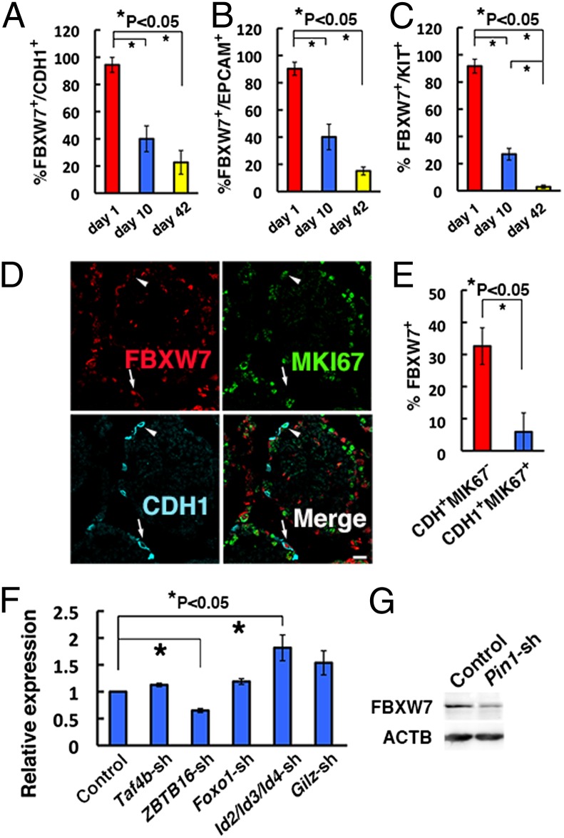
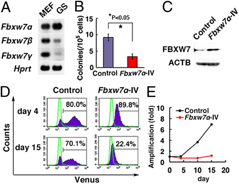
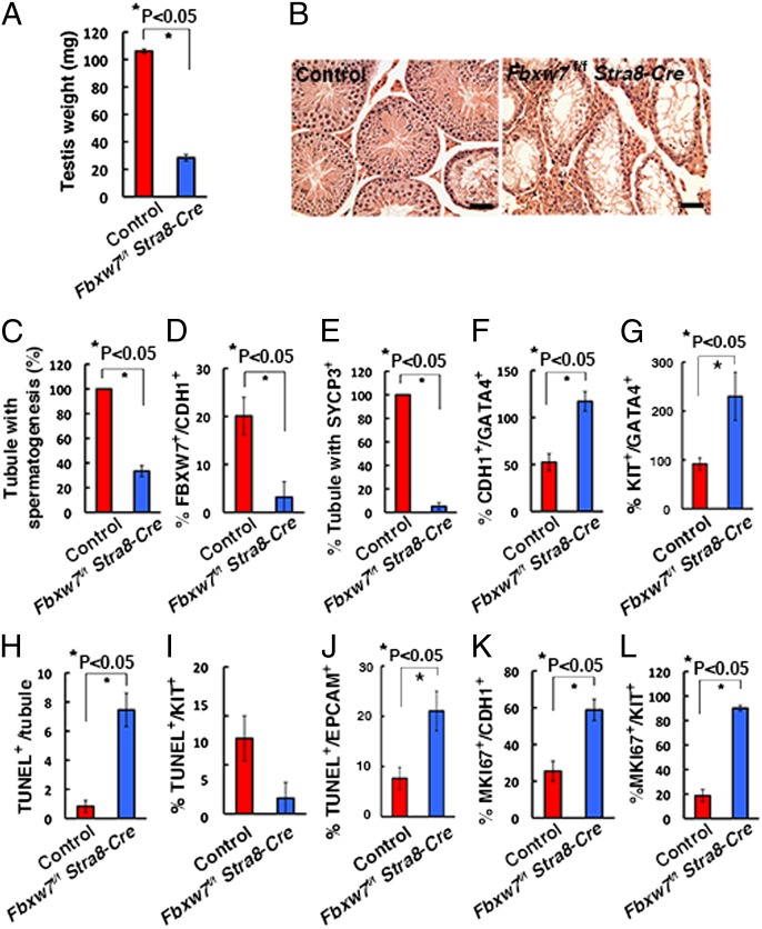
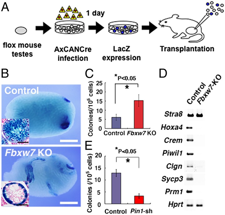
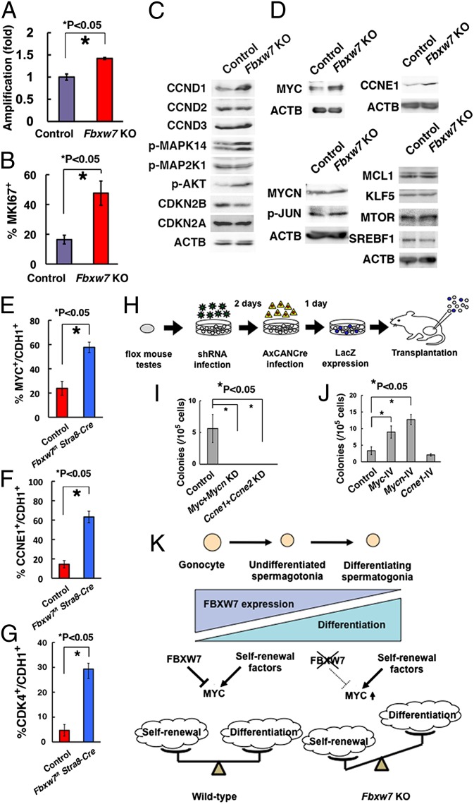
Spermatogonial stem cells (SSCs) undergo self-renewal divisions to support spermatogenesis throughout life. Although several positive regulators of SSC self-renewal have been discovered, little is known about the negative regulators. Here, we report that F-box and WD-40 domain protein 7 (FBXW7), a component of the Skp1-Cullin-F-box-type ubiquitin ligase, is a negative regulator of SSC self-renewal. FBXW7 is expressed in undifferentiated spermatogonia in a cell cycle-dependent manner. Although peptidyl-prolyl cis/trans isomerase NIMA-interacting 1 (PIN1), essential for spermatogenesis, is thought to destroy FBXW7, Pin1 depletion decreased FBXW7 expression. Spermatogonial transplantation showed that Fbxw7 overexpression compromised SSC activity whereas Fbxw7 deficiency enhanced SSC colonization and caused accumulation of undifferentiated spermatogonia, suggesting that the level of FBXW7 is critical for self-renewal and differentiation. Screening of putative FBXW7 targets revealed that Fbxw7 deficiency up-regulated myelocytomatosis oncogene (MYC) and cyclin E1 (CCNE1). Although depletion of Myc/Mycn or Ccne1/Ccne2 compromised SSC activity, overexpression of Myc, but not Ccne1, increased colonization of SSCs. These results suggest that FBXW7 regulates SSC self-renewal in a negative manner by degradation of MYC.
Conflict of interest statement
The authors declare no conflict of interest.
Figures
Similar articles
-
Fbxw7 contributes to tumor suppression by targeting multiple proteins for ubiquitin-dependent degradation.Cancer Sci. 2006 Aug;97(8):729-36. doi: 10.1111/j.1349-7006.2006.00239.x. Cancer Sci. 2006. PMID: 16863506 Free PMC article.
-
MicroRNA-223 regulates cyclin E activity by modulating expression of F-box and WD-40 domain protein 7.J Biol Chem. 2010 Nov 5;285(45):34439-46. doi: 10.1074/jbc.M110.152306. Epub 2010 Sep 7. J Biol Chem. 2010. PMID: 20826802 Free PMC article.
-
Myc/Mycn-mediated glycolysis enhances mouse spermatogonial stem cell self-renewal.Genes Dev. 2016 Dec 1;30(23):2637-2648. doi: 10.1101/gad.287045.116. Genes Dev. 2016. PMID: 28007786 Free PMC article.
-
Fbxw7 in cell cycle exit and stem cell maintenance: insight from gene-targeted mice.Cell Cycle. 2008 Nov 1;7(21):3307-13. doi: 10.4161/cc.7.21.6931. Epub 2008 Nov 5. Cell Cycle. 2008. PMID: 18948752 Review.
-
The Highs and Lows of FBXW7: New Insights into Substrate Affinity in Disease and Development.Cells. 2023 Aug 24;12(17):2141. doi: 10.3390/cells12172141. Cells. 2023. PMID: 37681873 Free PMC article. Review.
Cited by
-
Transfer of a Mouse Artificial Chromosome into Spermatogonial Stem Cells Generates Transchromosomic Mice.Stem Cell Reports. 2017 Oct 10;9(4):1180-1191. doi: 10.1016/j.stemcr.2017.08.012. Epub 2017 Sep 21. Stem Cell Reports. 2017. PMID: 28943251 Free PMC article.
-
Regulation of germ line stem cell homeostasis.Anim Reprod. 2015 Jan-Mar;12(1):35-45. Anim Reprod. 2015. PMID: 28286576 Free PMC article.
-
An interplay of NOX1-derived ROS and oxygen determines the spermatogonial stem cell self-renewal efficiency under hypoxia.Genes Dev. 2021 Feb 1;35(3-4):250-260. doi: 10.1101/gad.339903.120. Epub 2021 Jan 14. Genes Dev. 2021. PMID: 33446567 Free PMC article.
-
Targeting PD-1 post-translational modifications for improving cancer immunotherapy.Cell Insight. 2025 Apr 10;4(3):100248. doi: 10.1016/j.cellin.2025.100248. eCollection 2025 Jun. Cell Insight. 2025. PMID: 40336591 Free PMC article. Review.
-
Environmental pollutant Di-(2-ethylhexyl) phthalate induces asthenozoospermia: new insights from network toxicology.Mol Divers. 2025 Jun;29(3):2179-2192. doi: 10.1007/s11030-024-10976-9. Epub 2024 Sep 11. Mol Divers. 2025. PMID: 39259422
References
-
- de Rooij DG, Russell LD. All you wanted to know about spermatogonia but were afraid to ask. J Androl. 2000;21(6):776–798. - PubMed
-
- Meistrich ML, van Beek MEAB. Cell and molecular biology of the testis. In: Desjardins C, Ewing LL, editors. Spermatogonial Stem Cells. New York: Oxford Univ Press; 1993. pp. 266–295.
-
- Meng X, et al. Regulation of cell fate decision of undifferentiated spermatogonia by GDNF. Science. 2000;287(5457):1489–1493. - PubMed
-
- Tegelenbosch RAJ, de Rooij DG. A quantitative study of spermatogonial multiplication and stem cell renewal in the C3H/101 F1 hybrid mouse. Mutat Res. 1993;290(2):193–200. - PubMed
Publication types
MeSH terms
Substances
LinkOut - more resources
Full Text Sources
Other Literature Sources
Medical
Molecular Biology Databases
Research Materials
Miscellaneous
