

A versatile system for USER cloning-based assembly of expression vectors for mammalian cell engineering
- PMID: 24879460
- PMCID: PMC4039435
- DOI: 10.1371/journal.pone.0096693
A versatile system for USER cloning-based assembly of expression vectors for mammalian cell engineering
Abstract
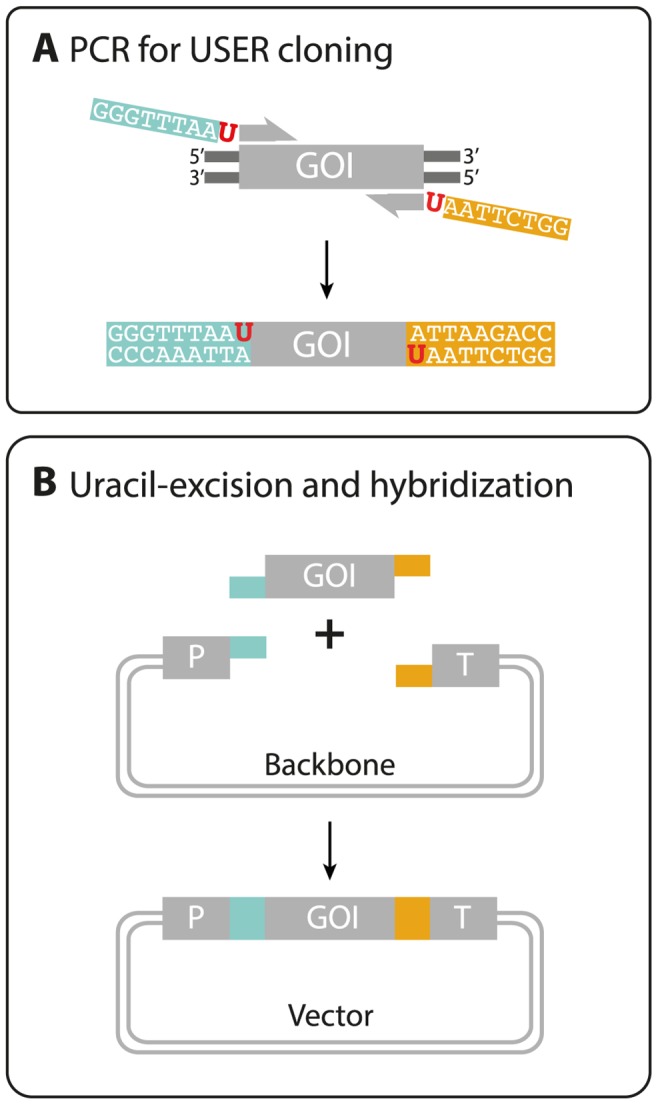
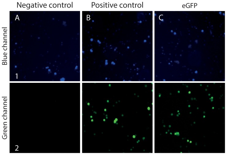
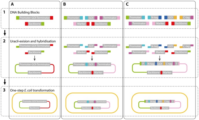
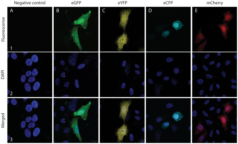
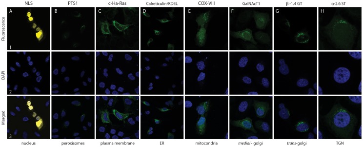
A new versatile mammalian vector system for protein production, cell biology analyses, and cell factory engineering was developed. The vector system applies the ligation-free uracil-excision based technique--USER cloning--to rapidly construct mammalian expression vectors of multiple DNA fragments and with maximum flexibility, both for choice of vector backbone and cargo. The vector system includes a set of basic vectors and a toolbox containing a multitude of DNA building blocks including promoters, terminators, selectable marker- and reporter genes, and sequences encoding an internal ribosome entry site, cellular localization signals and epitope- and purification tags. Building blocks in the toolbox can be easily combined as they contain defined and tested Flexible Assembly Sequence Tags, FASTs. USER cloning with FASTs allows rapid swaps of gene, promoter or selection marker in existing plasmids and simple construction of vectors encoding proteins, which are fused to fluorescence-, purification-, localization-, or epitope tags. The mammalian expression vector assembly platform currently allows for the assembly of up to seven fragments in a single cloning step with correct directionality and with a cloning efficiency above 90%. The functionality of basic vectors for FAST assembly was tested and validated by transient expression of fluorescent model proteins in CHO, U-2-OS and HEK293 cell lines. In this test, we included many of the most common vector elements for heterologous gene expression in mammalian cells, in addition the system is fully extendable by other users. The vector system is designed to facilitate high-throughput genome-scale studies of mammalian cells, such as the newly sequenced CHO cell lines, through the ability to rapidly generate high-fidelity assembly of customizable gene expression vectors.
Conflict of interest statement
Figures
Similar articles
-
A golden gate modular cloning toolbox for plants.ACS Synth Biol. 2014 Nov 21;3(11):839-43. doi: 10.1021/sb4001504. Epub 2014 Feb 20. ACS Synth Biol. 2014. PMID: 24933124
-
Construction of E. coli-Mycobacterium shuttle vectors with a variety of expression systems and polypeptide tags for gene expression in mycobacteria.PLoS One. 2020 Mar 11;15(3):e0230282. doi: 10.1371/journal.pone.0230282. eCollection 2020. PLoS One. 2020. PMID: 32160243 Free PMC article.
-
Construction of modular and versatile plasmid vectors for the high-level expression of single or multiple genes in insects and insect cell lines.J Mol Biol. 1999 Apr 23;288(1):13-20. doi: 10.1006/jmbi.1999.2674. J Mol Biol. 1999. PMID: 10329122
-
Phage N15-Based Vectors for Gene Cloning and Expression in Bacteria and Mammalian Cells.ACS Synth Biol. 2023 Apr 21;12(4):909-921. doi: 10.1021/acssynbio.2c00580. Epub 2023 Apr 6. ACS Synth Biol. 2023. PMID: 37026178 Review.
-
Designing plasmid vectors.Methods Mol Biol. 2009;542:117-29. doi: 10.1007/978-1-59745-561-9_6. Methods Mol Biol. 2009. PMID: 19565899 Review.
Cited by
-
Evolution-aided engineering of plant specialized metabolism.aBIOTECH. 2021 Jun 19;2(3):240-263. doi: 10.1007/s42994-021-00052-3. eCollection 2021 Sep. aBIOTECH. 2021. PMID: 36303885 Free PMC article. Review.
-
Continuous evolution of Bacillus thuringiensis toxins overcomes insect resistance.Nature. 2016 May 5;533(7601):58-63. doi: 10.1038/nature17938. Epub 2016 Apr 27. Nature. 2016. PMID: 27120167 Free PMC article.
-
Evaluating Apoptotic Gene Efficiency for CHO Culture Performance Using Targeted Integration.ACS Synth Biol. 2025 May 16;14(5):1414-1424. doi: 10.1021/acssynbio.4c00382. Epub 2025 Apr 23. ACS Synth Biol. 2025. PMID: 40268279 Free PMC article.
-
Versatile microscale screening platform for improving recombinant protein productivity in Chinese hamster ovary cells.Sci Rep. 2015 Dec 11;5:18016. doi: 10.1038/srep18016. Sci Rep. 2015. PMID: 26657798 Free PMC article.
-
A roadmap for gene system development in Clostridium.Anaerobe. 2016 Oct;41:104-112. doi: 10.1016/j.anaerobe.2016.05.011. Epub 2016 May 24. Anaerobe. 2016. PMID: 27234263 Free PMC article. Review.
References
-
- Walsh G (2010) Biopharmaceuticals benchmarks. Nat. Biotechnol. 28: 917–924. - PubMed
-
- Lewis NE, Liu X, Li Y, Nagarajan H, Yerganian G, et al. (2013) Genomic landscapes of Chinese hamster ovary cell lines as revealed by the Cricetulus griseus draft genome. Nat. Biotechnol 8: 759–65. - PubMed
-
- Wurm FM, Hacker D (2011) First CHO genome. Nat Biotechnol. 29: 718–20. - PubMed
-
- Ellis T, Adie T, Baldwin GS (2011) DNA assembly for synthetic biology: from parts to pathways and beyond. Integr. Biol. 3: 109–118. - PubMed
Publication types
MeSH terms
Substances
LinkOut - more resources
Full Text Sources
Other Literature Sources
Research Materials
