Review
. 2014 May 28;12(6):3161-89.
doi: 10.3390/md12063161.

Marennine, promising blue pigments from a widespread Haslea diatom species complex

Affiliations
Review

Marennine, promising blue pigments from a widespread Haslea diatom species complex

Romain Gastineau et al. Mar Drugs. .

Abstract

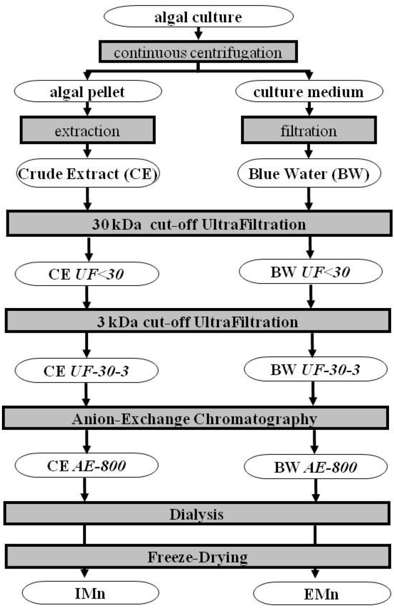
In diatoms, the main photosynthetic pigments are chlorophylls a and c, fucoxanthin, diadinoxanthin and diatoxanthin. The marine pennate diatom Haslea ostrearia has long been known for producing, in addition to these generic pigments, a water-soluble blue pigment, marennine. This pigment, responsible for the greening of oysters in western France, presents different biological activities: allelopathic, antioxidant, antibacterial, antiviral, and growth-inhibiting. A method to extract and purify marennine has been developed, but its chemical structure could hitherto not be resolved. For decades, H. ostrearia was the only organism known to produce marennine, and can be found worldwide. Our knowledge about H. ostrearia-like diatom biodiversity has recently been extended with the discovery of several new species of blue diatoms, the recently described H. karadagensis, H. silbo sp. inedit. and H. provincialis sp. inedit. These blue diatoms produce different marennine-like pigments, which belong to the same chemical family and present similar biological activities. Aside from being a potential source of natural blue pigments, H. ostrearia-like diatoms thus present a commercial potential for aquaculture, cosmetics, food and health industries.


Figures

Figure 1
Figure 1
Light micrograph of Haslea ostrearia.
Figure 2
Figure 2
Greening effect of marennine on bivalves. (a) Pacific oyster; (b) Scallop; (c) Cockle; (d) Clam.
Figure 3
Figure 3
Light micrograph of Haslea karadagensis.
Figure 4
Figure 4
Raman spectra obtained in vivo on the blue pigments contained in the apices of different strains of blue diatoms. Noticeable differences can be seen between the pigment of H. karadagensis and the others in the 1240 cm−1 to 1420 cm−1 region.
Figure 5
Figure 5
[1H NMR]. 1H 1D NMR spectra of the intracellular form (IMn) of marennine from Haslea ostrearia (bottom, green), the extracellular form (EMn) of marennine (middle, blue), and the extracellular form of the marennine-like pigment from Haslea provincialis sp. inedit. (top, red), all dissolved in D2O. The signal at 4.7 ppm stems from residual HDO, the high signal at 3.64 ppm in the extracellular marennine partly stems from an impurity.
Figure 6
Figure 6
Purification process of marennine and marennine-like pigments.
Figure 7
Figure 7
(a) Photography of the two PBRs used in this experiment; (b) Marennine concentration (mg L−1) in the PBR, measured spectrophotometrically on the culture medium. Values are means of concentrations obtained in each PBR (n = 2).
Figure 8
Figure 8
[2DNMR]. (a) 1H–13C HSQC of the extracellular form of marennine (EMn) in deuterated phosphate buffer. On top the corresponding 1D 1H spectrum. The lines separate areas of carbons with—among others—two hydrogen atoms and with one or three hydrogens, as determined by an additional edited HSQC with lower resolution and signal-to-noise ratio; (b) Generic pyranose; (c) Ring region of a 1H–1H TOCSY of EMn with 90 ms mixing time. The strong 1H signal at 3.65 ppm stems from an impurity.
Figure 9
Figure 9
Growth of Vibrio splendidus after 3 h in contact with marennine. V. splendidus was grown in modified marine media overnight. Cells were washed and then incubated for 3 h in 0, 0.1, 1.0, 10, 100, or 1000 μg mL−1 marennine. Cells were washed in sterile water and brought to an optical density of 0.5 before the 3 h incubation. Cells were then added to fresh marine media in a 96 well plate and growth kinetics were done for 48 h with measurements every 30 min. Inset: maximum growth rates expressed in function of the control (100%) with marennine concentrations.

References

    1. Wrede F., Strack E. Über das Pyocyanin, den blauen Farbstoff des Bacillus pyocyaneus. Z. Physiol. Chem. 1929;181:58–76. doi: 10.1515/bchm2.1929.181.1-3.58. - DOI
    1. Frank L.H., DeMoss R.D. On the biosynthesis of pyocyanine. J. Bacteriol. 1959;77:776–782. - PMC - PubMed
    1. Hassan H.M., Fridovich I. Mechanism of the antibiotic action of pyocyanine. J. Bacteriol. 1980;141:156–163. - PMC - PubMed
    1. Fujikawa H., Akimoto R. New Blue Pigment Produced by Pantoea agglomerans and Its Production Characteristics at Various Temperatures. Appl. Environ. Microbiol. 2011;77:172–178. doi: 10.1128/AEM.00264-10. - DOI - PMC - PubMed
    1. Fenical W. Chemical Studies of Marine Bacteria: Developing a New Resource. Chem. Rev. 1999;93:1673–1683. doi: 10.1021/cr00021a001. - DOI

Publication types