

Missing Concepts in De Novo Pulp Regeneration
- PMID: 24879576
- PMCID: PMC4126221
- DOI: 10.1177/0022034514537829
Missing Concepts in De Novo Pulp Regeneration
Abstract
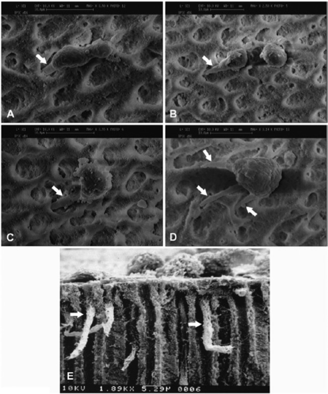
Regenerative endodontics has gained much attention in the past decade because it offers an alternative approach in treating endodontically involved teeth. Instead of filling the canal space with artificial materials, it attempts to fill the canal with vital tissues. The objective of regeneration is to regain the tissue and restore its function to the original state. In terms of pulp regeneration, a clinical protocol that intends to reestablish pulp/dentin tissues in the canal space has been developed--termed revitalization or revascularization. Histologic studies from animal and human teeth receiving revitalization have shown that pulp regeneration is difficult to achieve. In tissue engineering, there are 2 approaches to regeneration tissues: cell based and cell free. The former involves transplanting exogenous cells into the host, and the latter does not. Revitalization belongs to the latter approach. A number of crucial concepts have not been well discussed, noted, or understood in the field of regenerative endodontics in terms of pulp/dentin regeneration: (1) critical size defect of dentin and pulp, (2) cell lineage commitment to odontoblasts, (3) regeneration vs. repair, and (4) hurdles of cell-based pulp regeneration for clinical applications. This review article elaborates on these missing concepts and analyzes them at their cellular and molecular levels, which will in part explain why the non-cell-based revitalization procedure is difficult to establish pulp/dentin regeneration. Although the cell-based approach has been proven to regenerate pulp/dentin, such an approach will face barriers--with the key hurdle being the shortage of the current good manufacturing practice facilities, discussed herein.
Keywords: cell free; cell-based therapy; critical size defect; dentin; endodontics; stem cells.
© International & American Associations for Dental Research.
Conflict of interest statement
The authors declare no potential conflicts of interest with respect to the authorship and/or publication of this article.
Figures
Similar articles
-
Pulp development, repair, and regeneration: challenges of the transition from traditional dentistry to biologically based therapies.J Endod. 2014 Apr;40(4 Suppl):S2-5. doi: 10.1016/j.joen.2014.01.018. J Endod. 2014. PMID: 24698689
-
Dental pulp and dentin tissue engineering and regeneration: advancement and challenge.Front Biosci (Elite Ed). 2011 Jan 1;3(2):788-800. doi: 10.2741/e286. Front Biosci (Elite Ed). 2011. PMID: 21196351 Free PMC article. Review.
-
Regenerative endodontics: regeneration or repair?J Endod. 2014 Apr;40(4 Suppl):S70-5. doi: 10.1016/j.joen.2014.01.024. J Endod. 2014. PMID: 24698698
-
The coming era of regenerative endodontics: what an endodontist needs to know.Alpha Omegan. 2011 Spring;104(1-2):46-51. Alpha Omegan. 2011. PMID: 21905366
-
Pulp Regeneration Concepts for Nonvital Teeth: From Tissue Engineering to Clinical Approaches.Tissue Eng Part B Rev. 2018 Dec;24(6):419-442. doi: 10.1089/ten.TEB.2018.0073. Epub 2018 Jun 25. Tissue Eng Part B Rev. 2018. PMID: 29724156 Review.
Cited by
-
Potential of tailored amorphous multiporous calcium silicate glass for pulp capping regenerative endodontics-A preliminary assessment.J Dent. 2021 Jun;109:103655. doi: 10.1016/j.jdent.2021.103655. Epub 2021 Mar 30. J Dent. 2021. PMID: 33798640 Free PMC article.
-
Antimicrobial potential of resin matrices loaded with coffee compounds.J Biomed Mater Res B Appl Biomater. 2021 Mar;109(3):428-435. doi: 10.1002/jbm.b.34711. Epub 2020 Aug 25. J Biomed Mater Res B Appl Biomater. 2021. PMID: 32964641 Free PMC article.
-
Retreatment of failed revascularization/revitalization of immature permanent tooth - A case report.J Clin Exp Dent. 2018 Feb 1;10(2):e185-e188. doi: 10.4317/jced.53745. eCollection 2018 Feb. J Clin Exp Dent. 2018. PMID: 29670738 Free PMC article.
-
Regenerative Dentistry: Animal Model for Regenerative Endodontology.Transfus Med Hemother. 2016 Sep;43(5):359-364. doi: 10.1159/000447644. Epub 2016 Sep 6. Transfus Med Hemother. 2016. PMID: 27790081 Free PMC article.
-
Antibiofilm and immunomodulatory resorbable nanofibrous filing for dental pulp regenerative procedures.Bioact Mater. 2022 Feb 1;16:173-186. doi: 10.1016/j.bioactmat.2022.01.027. eCollection 2022 Oct. Bioact Mater. 2022. PMID: 35386316 Free PMC article.
References
-
- Angtrakool P. (2005). International standard ISO 14644 cleanrooms and associated controlled environments. Washington, DC: Food and Drug Administration, pp. 1-84. URL accessed on 5/9/2014 at: http://drug.fda.moph.go.th/drug/zone_gmp/files/GMP2549_2/Aug2106/7.ISO14....
-
- Bouillaguet S. (2004). Biological risks of resin-based materials to the dentin-pulp complex. Crit Rev Oral Biol Med 15:47-60. - PubMed
-
- Chadipiralla K, Yochim JM, Bahuleyan B, Huang CY, Garcia-Godoy F, Murray PE, et al. (2010). Osteogenic differentiation of stem cells derived from human periodontal ligaments and pulp of human exfoliated deciduous teeth. Cell Tissue Res 340:323-333. - PubMed
-
- Cowan CM, Shi YY, Aalami OO, Chou YF, Mari C, Thomas R, et al. (2004). Adipose-derived adult stromal cells heal critical-size mouse calvarial defects. Nat Biotechnol 22:560-567. - PubMed
-
- da Silva Meirelles L, Caplan AI, Nardi NB. (2008). In search of the in vivo identity of mesenchymal stem cells. Stem Cells 26:2287-2299. - PubMed
Publication types
MeSH terms
Grants and funding
LinkOut - more resources
Full Text Sources
Other Literature Sources
Miscellaneous
