

Ultrastructural abnormalities of the trabecular meshwork extracellular matrix in Cyp1b1-deficient mice
- PMID: 24879660
- PMCID: PMC4285769
- DOI: 10.1177/0300985814535613
Ultrastructural abnormalities of the trabecular meshwork extracellular matrix in Cyp1b1-deficient mice
Abstract
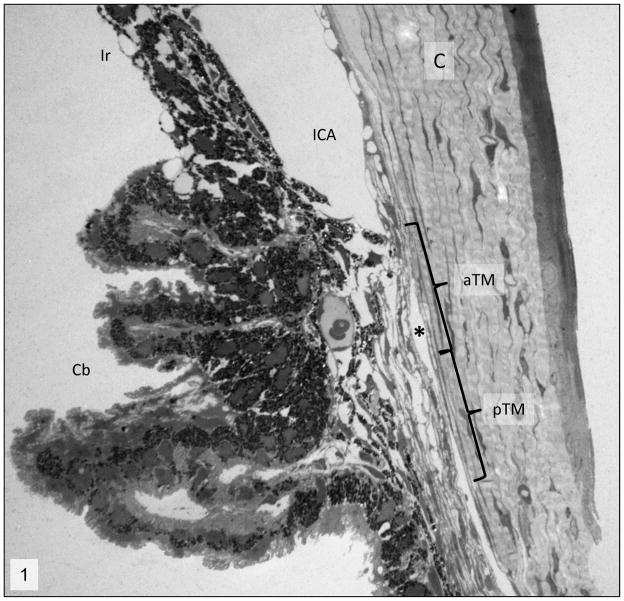
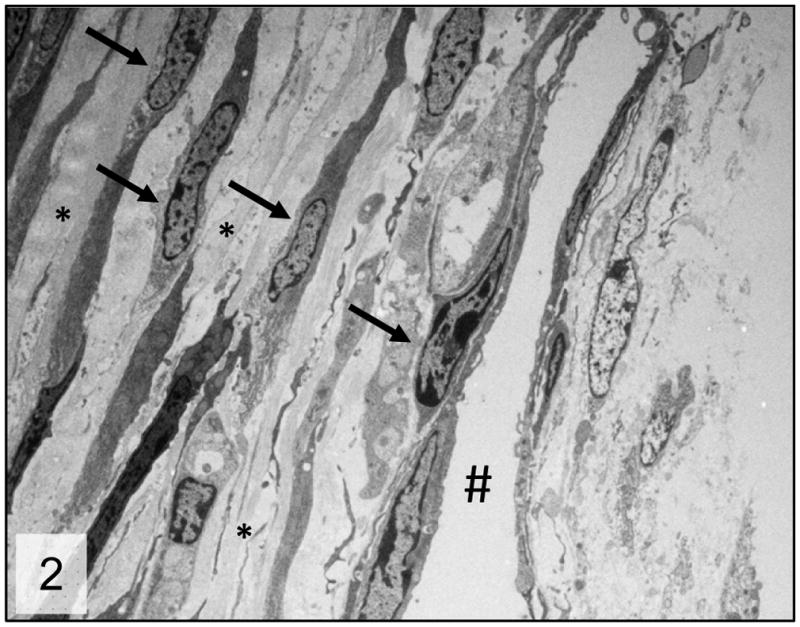
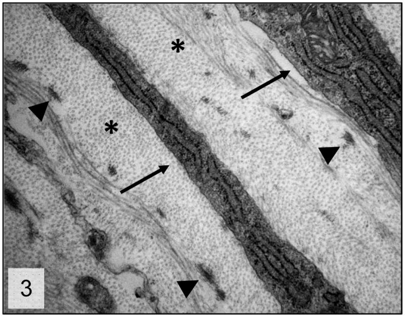
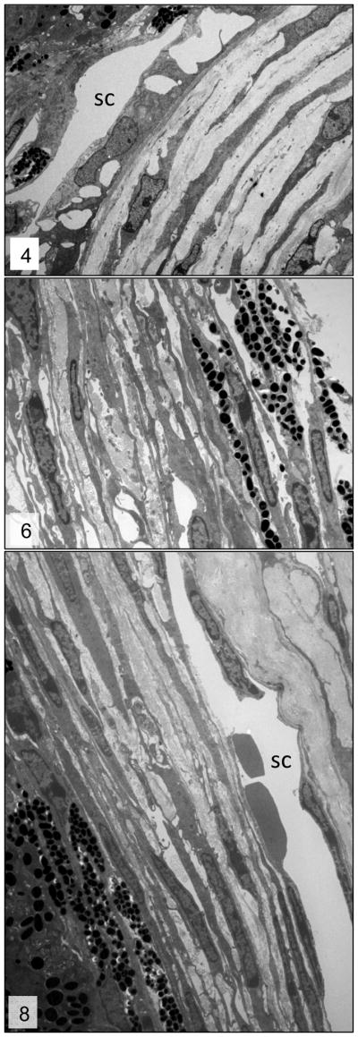
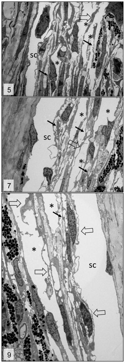
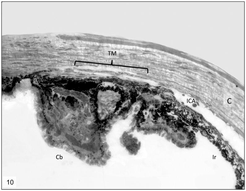
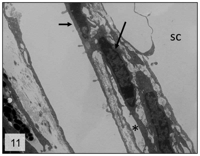
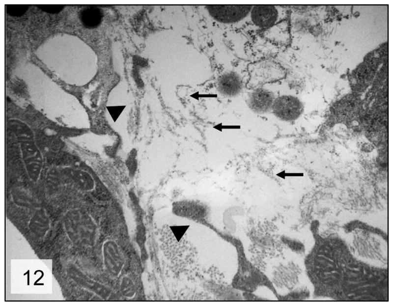
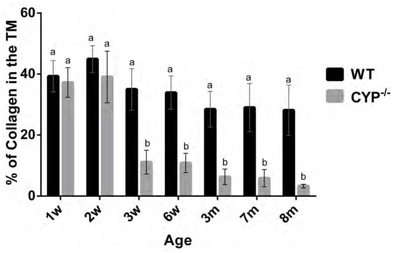
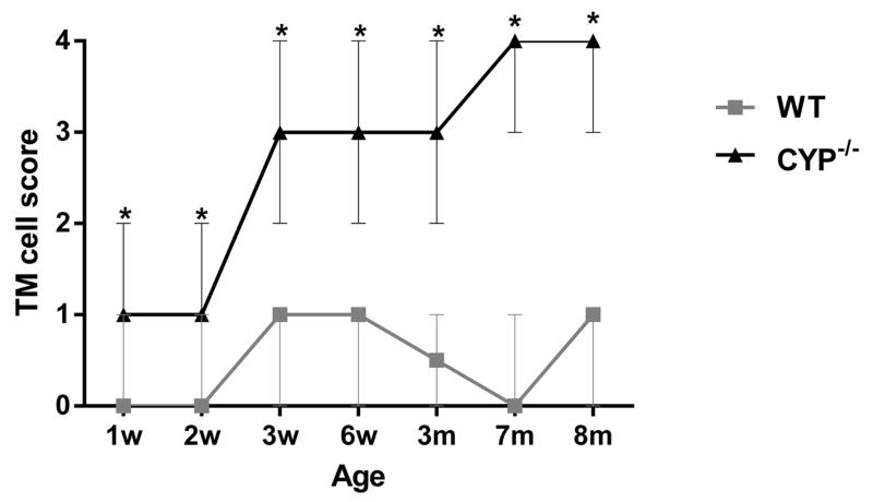
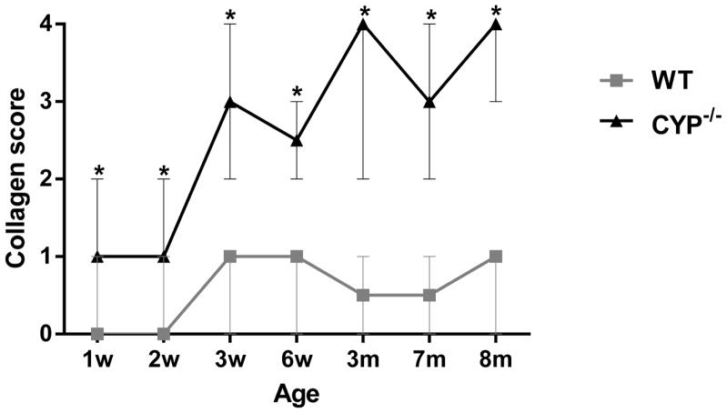
Cytochrome P450 1B1 (CYP1B1) is highly expressed in human and murine ocular tissues during development. Mutations in this gene are implicated in the development of primary congenital glaucoma (PCG) in humans. Mice deficient in Cyp1b1 (Cyp1b1(-/-) ) present developmental abnormalities similar to human primary congenital glaucoma. The present work describes the ultrastructural morphology of the iridocorneal angle of 21 eyes from 1-week-old to 8-month-old Cyp1b1(-/-) mice. Morphometric and semiquantitative analysis of the data revealed that 3-week-old Cyp1b1(-/-) mice present a significantly (P < .005) decreased amount of trabecular meshwork (TM) collagen and higher TM endothelial cell and collagen lesion scores (P < .005) than age-matched controls. Collagen loss and lesion scores were progressively increased in older animals, with 8-month-old animals presenting severe atrophy of the TM. Our findings advance the understanding of the effects of CYP1B1 mutations in TM development and primary congenital glaucoma, as well as suggest a link between TM morphologic alterations and increased intraocular pressure.
Keywords: CYP1B1; collagen fibrils; congenital glaucoma; electron microscopy; eye; mouse; oxidative stress; trabecular meshwork.
© The Author(s) 2014.
Figures
Similar articles
-
Cyp1b1 mediates periostin regulation of trabecular meshwork development by suppression of oxidative stress.Mol Cell Biol. 2013 Nov;33(21):4225-40. doi: 10.1128/MCB.00856-13. Epub 2013 Aug 26. Mol Cell Biol. 2013. PMID: 23979599 Free PMC article.
-
The p.Gly61Glu Mutation in CYP1B1 Affects the Extracellular Matrix in Glaucoma Patients.Ophthalmic Res. 2016 Jul;56(2):98-103. doi: 10.1159/000443508. Epub 2016 Mar 17. Ophthalmic Res. 2016. PMID: 26982174
-
Genotype and phenotype correlations in congenital glaucoma.Trans Am Ophthalmol Soc. 2006;104:183-95. Trans Am Ophthalmol Soc. 2006. PMID: 17471339 Free PMC article.
-
Cytochrome P450 1B1 and Primary Congenital Glaucoma.J Ophthalmic Vis Res. 2015 Jan-Mar;10(1):60-7. doi: 10.4103/2008-322X.156116. J Ophthalmic Vis Res. 2015. PMID: 26005555 Free PMC article. Review.
-
CYP1B1-mediated Pathobiology of Primary Congenital Glaucoma.J Curr Glaucoma Pract. 2015 Sep-Dec;9(3):77-80. doi: 10.5005/jp-journals-10008-1189. Epub 2016 Feb 2. J Curr Glaucoma Pract. 2015. PMID: 26997841 Free PMC article. Review.
Cited by
-
Therapeutic Drugs and Devices for Tackling Ocular Hypertension and Glaucoma, and Need for Neuroprotection and Cytoprotective Therapies.Front Pharmacol. 2021 Sep 17;12:729249. doi: 10.3389/fphar.2021.729249. eCollection 2021. Front Pharmacol. 2021. PMID: 34603044 Free PMC article. Review.
-
Null cyp1b1 Activity in Zebrafish Leads to Variable Craniofacial Defects Associated with Altered Expression of Extracellular Matrix and Lipid Metabolism Genes.Int J Mol Sci. 2021 Jun 16;22(12):6430. doi: 10.3390/ijms22126430. Int J Mol Sci. 2021. PMID: 34208498 Free PMC article.
-
Goniodysgenesis variability and activity of CYP1B1 genotypes in primary congenital glaucoma.PLoS One. 2017 Apr 27;12(4):e0176386. doi: 10.1371/journal.pone.0176386. eCollection 2017. PLoS One. 2017. PMID: 28448622 Free PMC article.
-
Challenges and Opportunities in P450 Research on the Eye.Drug Metab Dispos. 2023 Oct;51(10):1295-1307. doi: 10.1124/dmd.122.001072. Epub 2023 Mar 13. Drug Metab Dispos. 2023. PMID: 36914277 Free PMC article. Review.
-
Insights into CYP1B1-Related Ocular Diseases Through Genetics and Animal Studies.Life (Basel). 2025 Mar 3;15(3):395. doi: 10.3390/life15030395. Life (Basel). 2025. PMID: 40141740 Free PMC article. Review.
References
-
- Teixeira LBC, Dubielzig RR. The eye. In: Haschek WM, Rousseaux CG, Walling MA, editors. Haschek and Rousseaux’s Handbook of Toxicologic Pathology. 3. London, UK: Academic press; 2013. pp. 2095–2185.
-
- Weinreb RN, Khaw PT. Primary open-angle glaucoma. Lancet. 2004;363:1711–1720. - PubMed
-
- Tamm ER. The trabecular meshwork outflow pathways: structural and functional aspects. Exp Eye Res. 2009;88:648–655. - PubMed
-
- Panicker SG, Reddy AB, Mandal AK, et al. Identification of novel mutations causing familial primary congenital glaucoma in Indian pedigrees. Invest Ophthalmol Vis Sci. 2002;43:1358–1366. - PubMed
-
- Soley GC, Bosse KA, Flikier D, et al. Primary congenital glaucoma: a novel single-nucleotide deletion and varying phenotypic expression for the 1,546-1,555dup mutation in the GLC3A (CYP1B1) gene in 2 families of different ethnic origin. J Glaucoma. 2003;12:27–30. - PubMed
Publication types
MeSH terms
Substances
Grants and funding
LinkOut - more resources
Full Text Sources
Other Literature Sources
Medical
