Clinical Trial
. 2014 Jul 1;193(1):102-10.
doi: 10.4049/jimmunol.1400105. Epub 2014 May 30.

Hemin controls T cell polarization in sickle cell alloimmunization

Affiliations
Clinical Trial

Hemin controls T cell polarization in sickle cell alloimmunization

Hui Zhong et al. J Immunol. .

Abstract

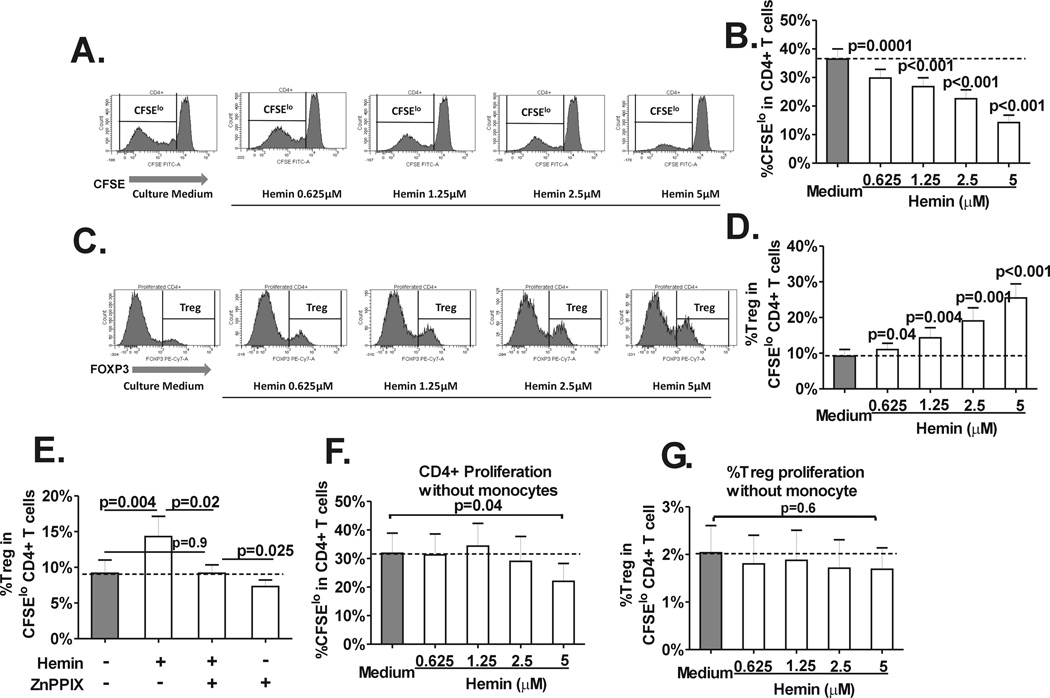
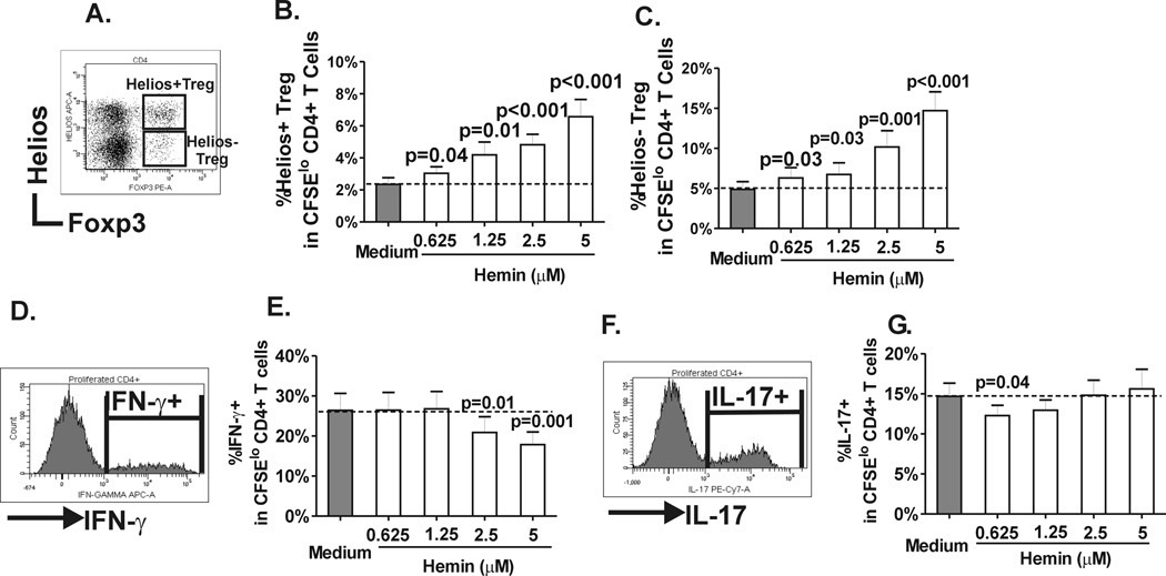
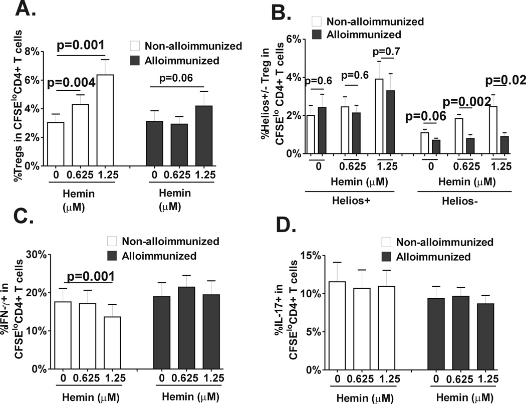
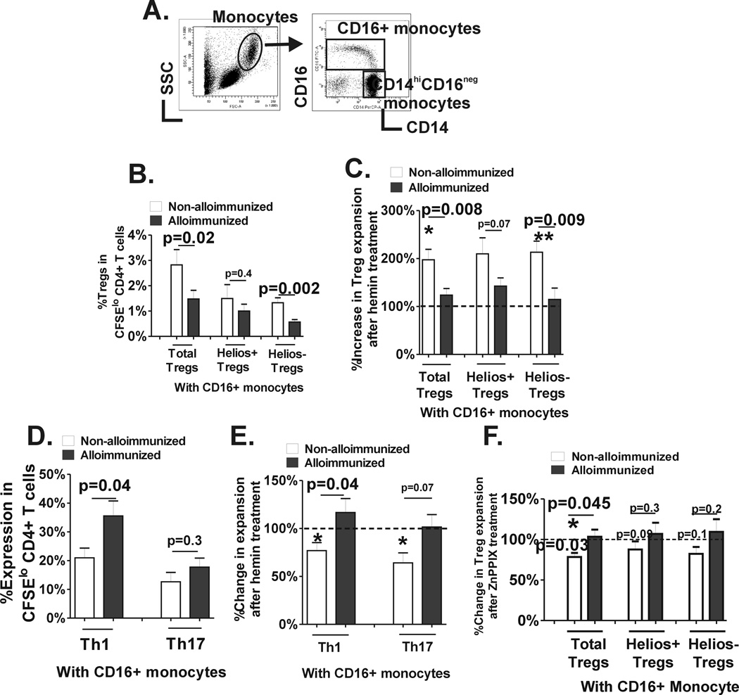
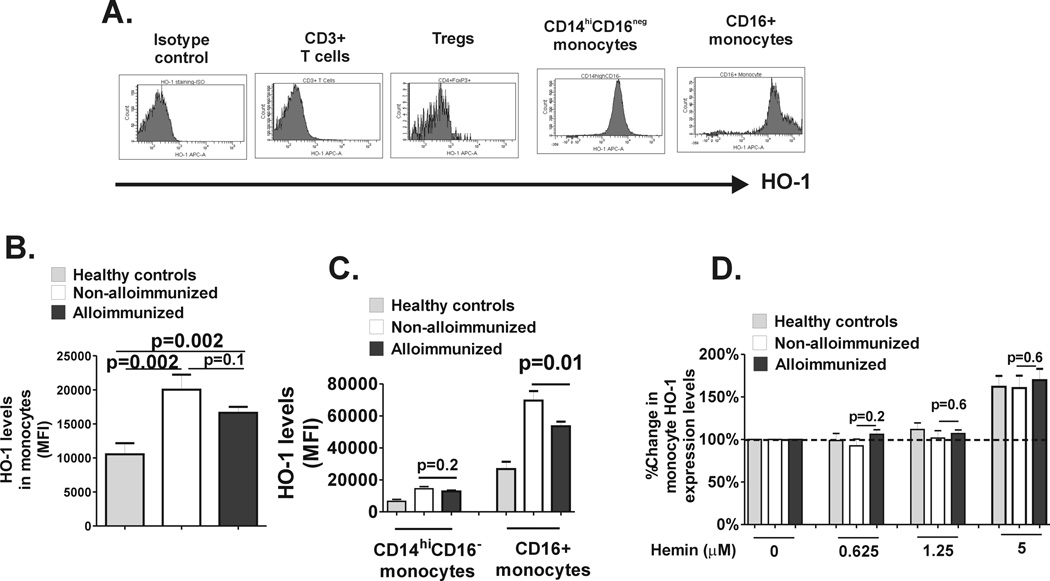
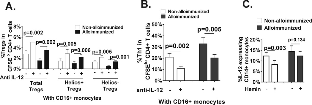
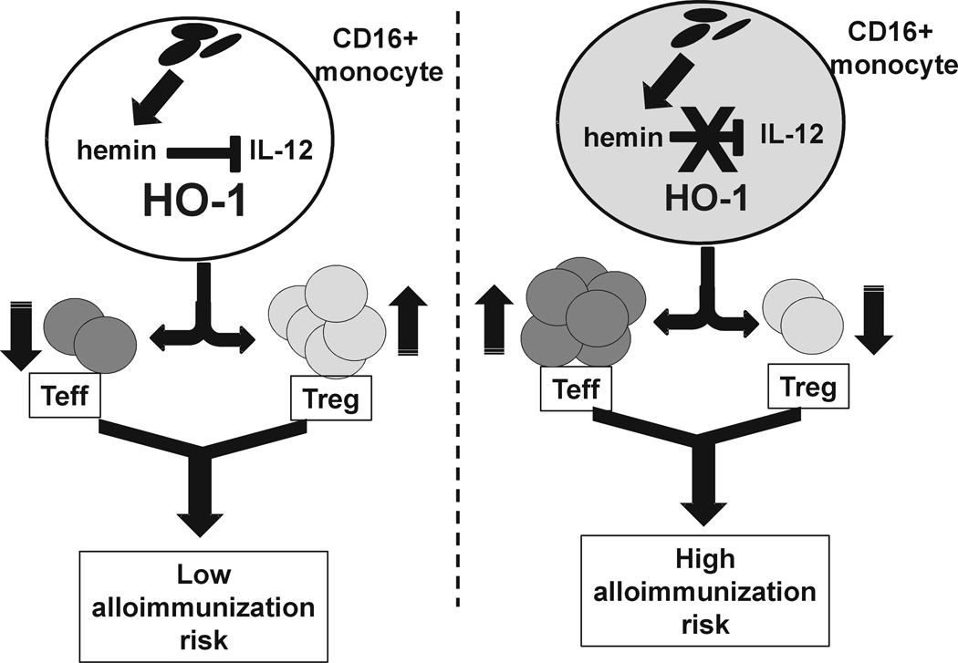
Patients with sickle cell disease (SCD) often require transfusions to treat and prevent worsening anemia and other SCD complications. However, transfusions can trigger alloimmunization against transfused RBCs with serious clinical sequelae. Risk factors for alloimmunization in SCD remain poorly understood. We recently reported altered regulatory T cell (Treg) and Th responses with higher circulating Th1 (IFN-γ(+)) cytokines in chronically transfused SCD patients with alloantibodies as compared with those without alloantibodies. Because monocytes play a critical role in polarization of T cell subsets and participate in clearance of transfused RBCs, we tested the hypothesis that in response to the RBC breakdown product hemin, monocyte control of T cell polarization will differ between alloimmunized and non-alloimmunized SCD patients. Exogenous hemin induced Treg polarization in purified T cell/monocyte cocultures from healthy volunteers through the monocyte anti-inflammatory heme-degrading enzyme heme oxygenase-1. Importantly, hemin primarily through its effect on CD16+ monocytes induced an anti-inflammatory (higher Treg/lower Th1) polarization state in the non-alloimmunized SCD group, whereas it had little effect in the alloimmunized group. Non-alloimmunized SCD CD16+ monocytes expressed higher basal levels of heme oxygenase-1. Furthermore, IL-12, which contributed to a proinflammatory polarization state (low Treg/high Th1) in SCD, was dampened in hemin-treated stimulated monocytes from non-alloimmunized SCD patients, but not in the alloimmunized group. These data suggest that unlike alloimmunized patients, non-alloimmunized SCD CD16+ monocytes in response to transfused RBC breakdown products promote an anti-inflammatory state that is less conducive to alloimmunization.


Figures

Fig. 1
Fig. 1. Hemin induces CD4+ Treg polarization
Purified, CFSE-stained CD4+ T cells from normal healthy volunteers (n=14) were co-cultured with autologous purified total monocyte fraction in the absence (“Culture medium”) or presence of various concentrations of hemin (0 to 5µM) and stimulated with anti-CD3 antibody for 7 days. (A) Representative histograms of the staining pattern of CFSE in total CD4+ T cells after 7 days in cocultures showing the gating used to measure the extent of CD4+ T cell proliferation defined as the frequency of divided (CFSElo) population. (B) Dose-dependent inhibitory effect of hemin on the frequency of CD4+ T cell proliferation (CD4+CFSElo cells) in T cell-monocyte cocultures from healthy controls. P values were analyzed by paired t test comparing before (“medium”) and after addition of hemin. (C) Representative histograms of the staining pattern of T cell subset expressing Foxp3hi (Tregs) within the CD4+CFSElo population in cocultures untreated or treated with various concentrations of hemin (0 to 5µM) showing the gating used to analyze the frequency of Treg population that had undergone proliferation. (D) Dose dependent increase in the frequency of Tregs in divided CD4+ T cells in the same (C) cocultures. (E) Frequency of divided Tregs in cocultures without or with addition of hemin (1.25µM) and/or HO-1 inhibitor ZnPPIX (2.5 µM) at day 0. The dotted line marks the basal Treg proliferation (minus hemin or ZnPPIX). Frequency of (F) total CD4+ and (G) Treg proliferation in cocultures treated with different doses of hemin in the absence of monocytes are shown. Unlike cocultures with monocytes, Treg proliferation is not affected by hemin and total CD4+ proliferation is only inhibited at the highest hemin concentration (5 µM). All statistical analysis indicated by p values was performed by paired t test.
Fig. 2
Fig. 2. Helios+/− Treg subset, Th1 and Th17 expansion in response to hemin
(A) Representative dot plot showing helios and Foxp3 staining pattern in divided (CFSElo) CD4+ T cells in T cell-monocyte cocultures on day 7. Helios+ Tregs are defined by co-expression of helios in Foxp3hi population whereas Helios− Tregs lack helios expression. Frequency of (B) helios+ and (C) helios− Tregs that had undergone proliferation in the same cocultures from healthy control as in Fig. 1 before and after hemin treatment. (C) Representative histogram of IFN-γ expression in divided (CFSElo) CD4+ T cells on day 7 and (D) frequencies of IFN-γ + cells in divided CD4+ cells in the absence or presence of increasing concentrations of hemin in the same cocultures from healthy control as in Fig. 1. Similarly, (E) representative histogram of IL-17 expression in divided (CFSElo) CD4+ T cells on day 7 and (F) frequencies of IL-17+ cells in divided CD4+ cells without or with hemin in cocultures from Fig. 1. P values were analyzed by paired t test comparing before (“medium”) and after addition of hemin.
Fig. 3
Fig. 3. Differences in Treg/Th polarization in response to hemin between alloimmunized and non-alloimmunized SCD patients
Purified, CFSE-stained CD4+ T cells from regularly transfused non-alloimmunized (n=9, white bars) and alloimmunized (n=11, black bars) SCD patients were co-cultured with autologous purified total monocyte fraction in the absence or presence of 2 different concentrations of hemin (0.625µM and 1.25µM) and stimulated with anti-CD3 antibody for 7 days. Levels of (A) total Tregs, (B) helios+/− Treg subsets, (C) Th1 and (D) Th17 in CD4+ T population that had undergone proliferation were analyzed by flow cytometry. All statistical analysis comparing before and after hemin addition was performed using paired t test; comparison between alloimmunized and non-alloimmunized groups was performed using Mann-Whitney test.
Fig. 4
Fig. 4. CD16+ monocyte control of Treg/Th proliferation before and after hemin treatment
(A) Representative dot plot analysis of PBMCs based on forward and side scatter showing the gating strategy to identify the total monocyte population. Based on CD14 and CD16 expression pattern, CD16+ monocytes are further distinguished from CD14hiCD16 monocyte subset. Purified CFSE stained CD4+ T cells from non-alloimmunized (n=9, white bars) and alloimmunized (n=11, black bars) SCD patients were cocultured with autologous purified CD14+CD16 monocyte subset together with autologous purified CD16+ monocytes followed by stimulation with anti-CD3 for 7 days in the presence or absence of 1.25µM hemin. (B) Frequency of total and Helios helios +/− Treg subsets that had undergone proliferation in the absence of hemin are shown. (C) Fold change in proliferation of total and Helios helios+/− Treg subsets after addition of hemin was calculated (proliferation in the absence of hemin was set at 100%). The asterisks correspond to statistically significant differences in the proliferative responses before and after hemin treatment (paired t test). (D) Frequency IFN-γ+ and IL-17+ cells in divided CD4+ T cells from the same cocultures as in B are shown. (E) Fold change in Th1 and Th17 proliferation after addition of hemin was calculated (proliferation in the absence of hemin was set at 100%). The asterisks correspond to statistically significant differences in the proliferative responses before and after hemin treatment (paired t test). (F) Fold change in Treg proliferation after addition of ZnPPIX was calculated (proliferation in the absence of ZnPPIX was set at 100%). The p value indicated above the column was calculated by paired t test comparing before and after ZnPPIX treatment. All comparisons between alloimmunized and non-alloimmunized groups were performed using Mann-Whitney test.
Fig. 5
Fig. 5. HO-1 Expression levels of monocyte subset HO-1 levels
Levels of intracellular HO-1 expression were measured in PBMCs and (A) representative histograms showing isotype control and HO-1 expression in T cells (CD3+) and Tregs (CD25+Foxp3hi) as well as in CD14hiCD16 and CD16+ monocytes are shown. (B) HO-1 expression in total monocyte fraction and (C) in CD14hiCD16 and CD16+ monocyte sunsets in PBMCs from healthy controls (grey bars) and non-alloimmunized (white bars) and alloimmunized (black bars) SCD patient as measured by relative mean fluorescent intensity (MFI) are shown. (D) PBMCs from healthy controls and SCD patients were cultured without (0) or with various concentrations of hemin for 24 hours and fold change in HO-1 expression relative to no added hemin (set at 100%) was calculated. Although at lower concentrations of hemin (0.625µM and 1.25µM), there was no change in HO-1 levels, at higher concentration (5µM), HO-1 expression level was induced (more than 100%). The differences in HO-1 inducibility between the groups were not different at any of the hemin concentrations tested. All comparisons between alloimmunized and non-alloimmunized groups were performed using Mann-Whitney test.
Fig. 6
Fig. 6. Role of IL-12 in CD16+ monocyte control of Treg/Th polarization and response to hemin
Isotype control (“minus”) or neutralizing anti-IL12 p40/p70 (“plus”) was added to the T cell-monocytes cocultures that included CD16+ monocytes from non-alloimmunized (white bars, n=8) and alloimmunized (black bars, n=8) SCD patients at day 0 and frequencies of (A) total Tregs and Helios+/− Treg subsets and (B) IFN-γ+ cells in divided CD4+T cells was measured on day 7. All statistical analysis comparing isotype vs anti-IL-12 was performed using paired t test. (C) Frozen and then thawed PBMCs from non-alloimmunized (white bars, n=8) and alloimmunized (black bars, n=7) SCD patients were stimulated with IFN-γ in the absence or presence of hemin (1.25 µM) for 2 hours followed by addition of LPS for another 22 hours. Frequency of IL-12 expressing monocytes in untreated and hemin treated samples are shown and the p values indicate paired t-test comparison analysis of untreated vs hemin-treated samples.
Fig 7
Fig 7. Proposed mechanism of altered monocyte mediated Treg/Th polarization in SCD alloimmunization
Levels of hemin, a breakdown product of hemoglobin, are likely to build up in monocyte/macrophages following RBC transfusions. If proinflammatory cytokines including IL-12 are at low levels and are maintained at low levels in response to hemin due to adequate HO-1 in CD16+ monocytes, Treg/Th polarization will be switched toward a regulatory response (higher Treg/lower Th1) that is less conducive to alloimmunization. However, if IL-12 levels in CD16+ monocytes are not optimally inhibited by hemin as a result of insufficient HO-1 activity/level, Tregs will not expand and Th1 proliferation will dominate, thereby increasing the risk of alloimmunization.

Similar articles

Cited by

References

    1. Frenette PS, Atweh GF. Sickle cell disease: old discoveries, new concepts, and future promise. J. Clin. Invest. 2007;117:850–858. - PMC - PubMed
    1. Josephson CD, Su LL, Hillyer KL, Hillyer CD. Transfusion in the patient with sickle cell disease: a critical review of the literature and transfusion guidelines. Transfus. Med. Rev. 2007;21:118–133. - PubMed
    1. Yazdanbakhsh K, Ware RE, Noizat-Pirenne F. Red blood cell alloimmunization in sickle cell disease: pathophysiology, risk factors, and transfusion management. Blood. 2012;120:528–537. - PMC - PubMed
    1. Chou ST, Jackson T, Vege S, Smith-Whitley K, Friedman DF, Westhoff CM. High prevalence of red blood cell alloimmunization in sickle cell disease despite transfusion from Rh-matched minority donors. Blood. 2013;122:1062–1071. - PubMed
    1. Bao W, Zhong H, Li X, Lee MT, Schwartz J, Sheth S, Yazdanbakhsh K. Immune regulation in chronically transfused allo-antibody responder and nonresponder patients with sickle cell disease and beta-thalassemia major. Am. J. Hematol. 2011;86:1001–1006. - PMC - PubMed

Publication types

MeSH terms