. 2014 Jul;15(7):631-7.
doi: 10.1038/ni.2914. Epub 2014 Jun 1.

CD80 and PD-L2 define functionally distinct memory B cell subsets that are independent of antibody isotype

Affiliations

CD80 and PD-L2 define functionally distinct memory B cell subsets that are independent of antibody isotype

Griselda V Zuccarino-Catania et al. Nat Immunol. 2014 Jul.

Abstract

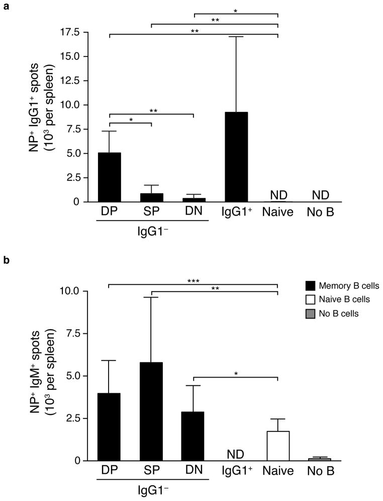
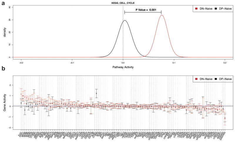
Memory B cells (MBCs) are long-lived sources of rapid, isotype-switched secondary antibody-forming cell (AFC) responses. Whether MBCs homogeneously retain the ability to self-renew and terminally differentiate or if these functions are compartmentalized into MBC subsets has remained unclear. It has been suggested that antibody isotype controls MBC differentiation upon restimulation. Here we demonstrate that subcategorizing MBCs on the basis of their expression of CD80 and PD-L2, independently of isotype, identified MBC subsets with distinct functions upon rechallenge. CD80(+)PD-L2(+) MBCs differentiated rapidly into AFCs but did not generate germinal centers (GCs); conversely, CD80(-)PD-L2(-) MBCs generated few early AFCs but robustly seeded GCs. The gene-expression patterns of the subsets supported both the identity and function of these distinct MBC types. Hence, the differentiation and regeneration of MBCs are compartmentalized.


Conflict of interest statement

COMPETING FINANCIAL INTERESTS

The authors declare no competing financial interests.

Figures

Figure 1
Figure 1
Generation and purification of MBC subsets. (a) Flow cytometry analysis of splenic cells from AM14 Tg x Vκ8R KI recipient mice that received NP-specific B cell and were at 8 weeks post-immunization with NP-CGG in alum. Left shows gating on antigen-specific B cells (CD19+NIP+) after gating on live cells and right shows staining for IgG1. Numbers indicate percentage of the parent-gated cells in the indicated population. Data are from one mouse from one experiment, representative of nine independent experiments with twenty to thirty mice per experiment. (b) Flow cytometry analysis of splenic B cells from AM14 Tg x Vκ8R KI recipient mice that did not receive NP-specific B cells but were immunized with NP-CGG in alum 8 weeks prior. Numbers indicate percentage of the parent-gated cells in the indicated population. Data are from one mouse from one experiment, representative of three independent experiments with three mice per experiment. (c) MBC subset distribution and frequency after gating as in (a), after staining for either IgG1 or IgM: (left) CD19+ NIP+ IgG1 MBCs (right) CD19+ NIP+ IgM MBCs. Subsets were identified according to expression of CD80 and PD-L2 that separates up to three populations: DP, SP and DN. Numbers indicate percentage of the parent gated cells in the indicated population. Data are from one mouse from one experiment, representative of nine independent experiments with twenty to thirty mice per experiment (left) or two independent experiments with forty-three to forty-five mice per experiment (right).
Figure 2
Figure 2
DP IgG1 and IgG1+ MBCs are the major producers of early IgG1+ AFCs. Numbers of AFCs per spleen were determined by ELISPOT after transfer of DP, SP or DN IgG1 MBCs; IgG1pos MBCs or naïve B cells in recipient mice 3.5 days post immunization with NP-OVA in alum. (a) Numbers of NP+ IgG1+ AFCs generated in mice transferred with the indicated populations of cells. ND, not detected. * p<0.001, ** p<0.0001. (Mann Whitney nonparametric, two-tailed test). Data are combined from two independent experiments (error bars represent standard deviation) with five to fourteen mice per group. (b) Numbers of IgM+ AFCs. ND, not detected. * p<0.05, ** p<0.001, *** p<0.0001. (Mann Whitney nonparametric, two-tailed test). Data are combined from two independent experiments (error bars represent standard deviation) with three to sixteen mice per group.
Figure 3
Figure 3
MBC requirement of T cells to generate early AFC responses. Numbers of AFCs per spleen were determined by ELISPOT analysis after transfer of DP, SP or DN IgG1 MBC; IgG1pos MBC or naïve B cells in recipient mice 3.5 days post immunization with NP-OVA in alum. Recipient mice were treated with anti-CD4 or PBS as a control before transfer of B cells. (a) Numbers of NP+ IgG1+ spleen AFCs. (b) Numbers of NP+ IgM+ spleen AFCs. Six to nineteen mice per group for PBS treated mice, and five to thirteen mice per group for anti-CD4 treated mice. ND, not detected. * p<0.05, ** p<0.01, *** p<0.001, **** p<0.0001. (Mann Whitney nonparametric, two-tailed test). Data are combined from two (for anti-CD4 treated mice) or three (for PBS treated mice) independent experiments (error bars represent standard deviation).
Figure 4
Figure 4
DP IgG1 MBCs promote more cognate T cell expansion than do other B cell types. Numbers of KJ1-26+ CD4+ OVA-specific T cells in mice that received memory T cells with DP, SP or DN IgG1 MBCs; IgG1+ MBCs; naïve B cell or no B cells, 3.5 days after immunization with NP-OVA in alum. * p<0.01, ** p<0.001, *** p<0.0001. (two-tailed t-test). Data are combined from two independent experiments. Error bars represent standard deviation, with six to sixteen mice per group.
Figure 5
Figure 5
GC B cells are generated from SP or DN IgG1 MBCs or naïve B cells, but not DP MBCs. Numbers of CD95+ CD38 NIP+ GC B cells 10.5 days after immunization generated after transfer of: (a) DP, SP or DN IgG1 MBCs; IgG1+ MBCs; naïve B cells or no B cells, along with memory T cells, or (b) DP or SP IgM MBCs, naive B cells or no B cells, along with memory T cells. ND, not detected. * p<0.05, ** p<0.01, *** p<0.0001. (two-tailed t-test). Data are combined from three independent experiments with eight to twenty-two mice per group for NP-OVA in alum immunized mice and one to fifteen mice per group for alum immunized mice (a), or two independent experiments with six to thirteen mice per group for NP-OVA in alum immunized mice or two to six mice per group for alum immunized mice (b). Error bars represent standard deviation.
Figure 6
Figure 6
Late IgG1+, but not IgM+, AFC formation is dominated by DN IgG1 MBCs and naïve B cells. Numbers of AFC per spleen were determined by ELISPOT analysis after transfer of DP, SP or DN IgG1 MBC; DP or SP IgM MBC; naïve B cell or no B cell responses in spleens of mice that received memory T cells 10.5 days post immunization with NP-OVA in alum or alum alone (not shown in a as there were no detectable responses). (a) Numbers of NP+ IgG1+ spleen AFCs. (b) Numbers of NP+ IgM+ spleen AFCs.* p<0.05, ** p<0.01, *** p<0.001, **** p<0.0001. (Mann Whitney nonparametric, two-tailed test). Data are combined from four independent experiments with seven to twenty-four mice per group (a) or two independent experiments with six to sixteen mice per group for alum immunized mice or one to eleven mice per group for alum immunized mice (b). ND, not detected. Error bars represent standard deviation.
Figure 7
Figure 7
The cell cycle pathway is significantly activated in the DN subset. QuSAGE was used to quantify the activity of the KEGG CELL CYCLE gene set. Data are derived from three biological replicates per cell type. (a) Activity Probability Density Functions (PDFs) of the DN subset relative to naïve B cells (red) and DP subset relative to naïve B cells (black). (b) Activity of individual genes in the KEGG CELL CYCLE pathway in the DN subset (red) and DP subset (black). Activity is quantified by the log2 fold-change relative to naïve B cells. Points indicate the mean and bars show the 95% confidence intervals.

Similar articles

Cited by

References

    1. Ahmed R, Gray D. Immunological memory and protective immunity: understanding their relation. Science. 1996;272:54–60. - PubMed
    1. Sallusto F, Lanzavecchia A, Araki K, Ahmed R. From vaccines to memory and back. Immunity. 2010;33:451–463. - PMC - PubMed
    1. Radbruch A, et al. Competence and competition: the challenge of becoming a long-lived plasma cell. Nat Rev Immunol. 2006;6:741–750. - PubMed
    1. McKinstry KK, et al. Rapid default transition of CD4 T cell effectors to functional memory cells. J Exp Med. 2007;204:2199–2211. - PMC - PubMed
    1. Bevan MJ. Understand memory, design better vaccines. Nat Immunol. 2011;12:463–465. - PMC - PubMed

Publication types

Substances

Associated data