. 2014 Aug;35(25):6941-53.
doi: 10.1016/j.biomaterials.2014.05.013. Epub 2014 May 29.

Silk as a biocohesive sacrificial binder in the fabrication of hydroxyapatite load bearing scaffolds

Affiliations

Silk as a biocohesive sacrificial binder in the fabrication of hydroxyapatite load bearing scaffolds

Stephanie L McNamara et al. Biomaterials. 2014 Aug.

Abstract

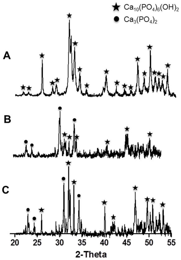
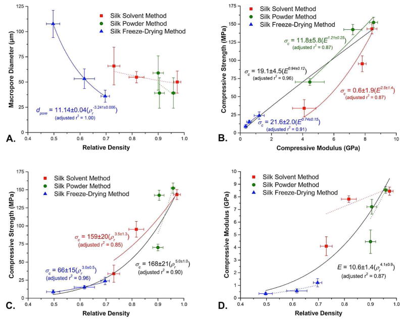
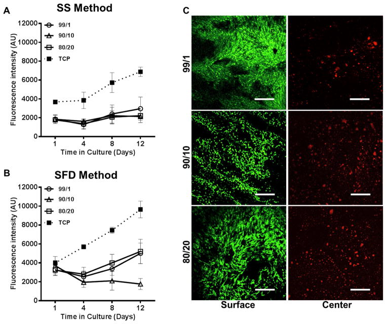
Limitations of current clinical methods for bone repair continue to fuel the demand for a high strength, bioactive bone replacement material. Recent attempts to produce porous scaffolds for bone regeneration have been limited by the intrinsic weakness associated with high porosity materials. In this study, ceramic scaffold fabrication techniques for potential use in load-bearing bone repairs have been developed using naturally derived silk from Bombyx mori. Silk was first employed for ceramic grain consolidation during green body formation, and later as a sacrificial polymer to impart porosity during sintering. These techniques allowed preparation of hydroxyapatite (HA) scaffolds that exhibited a wide range of mechanical and porosity profiles, with some displaying unusually high compressive strength up to 152.4 ± 9.1 MPa. Results showed that the scaffolds exhibited a wide range of compressive strengths and moduli (8.7 ± 2.7 MPa to 152.4 ± 9.1 MPa and 0.3 ± 0.1 GPa to 8.6 ± 0.3 GPa) with total porosities of up to 62.9 ± 2.7% depending on the parameters used for fabrication. Moreover, HA-silk scaffolds could be molded into large, complex shapes, and further machined post-sinter to generate specific three-dimensional geometries. Scaffolds supported bone marrow-derived mesenchymal stem cell attachment and proliferation, with no signs of cytotoxicity. Therefore, silk-fabricated HA scaffolds show promise for load bearing bone repair and regeneration needs.

Keywords: Bone tissue engineering; Ceramic structure; Hydroxyapatite; Porosity; Silk.


Figures

Figure 1
Figure 1
Silk-based processing methods for fabrication of HA scaffolds. A. Silk protein is used in the form of a concentrated aqueous solution in the Silk Solvent (SS) and Silk Freeze Drying (SFD) Methods or as a soluble silk powder, made by freeze-drying silk solution followed by blending, in the Silk Powder (SP) Method to bind ceramic particles together into complex geometry green bodies (B–D). The silk later acts as a sacrificial polymer during sintering to induce porosity in the final scaffolds (E), which can then be machined into any three-dimensional shape using standard machining techniques (F). Scale bars are 10 mm in B and E, and 5 mm in C, D, and F.
Figure 2
Figure 2
Shrinkage of silk-fabricated HA scaffolds during sintering. Shrinking of HA-silk green bodies during sintering at 1300°C was associated with a loss of both volume and mass as shown for green bodies with varying silk content (1% to 20% by mass) for A) Silk Powder Method, B) Silk Solvent Method, and C) Silk Freeze Drying (***p<0.001 and n.s. p>0.05, n = 10). Differences in mass loss with consistent decreases in volume for the three HA/silk ratios were observed, suggesting differences in final scaffold porosity.
Figure 3
Figure 3
Phase analysis of HA material and sintered silk-fabricated HA scaffolds. a) X-ray diffraction patterns of HA powder used for scaffold fabrication confirmed the presence of a single phase of HA and no trace of secondary phases or contaminating residues. b) XRD patterns of the sintered scaffold surface and c) sintered scaffold center confirmed the complete removal of silk protein during sintering based on the absence of characteristic silk peaks, and also revealed biphasic composition of the post-sintered material containing a mixture of both HA and alpha-tricalcium phosphate. Phase compositions were similar for scaffolds sintered at either 1300°C or 1400°C. A star denotes a peak corresponding to HA, and a circle denotes a peak corresponding to alpha-TCP.
Figure 4
Figure 4
Silk-fabricated HA scaffold morphology and porosity. a) Micro-computed tomography imaging at 25X magnification of both the surface and cross-section of silk-fabricated HA scaffolds made by SP, SS, and SFD methods with varying silk content (1% to 20% by mass) and sintered at 1300°C. Scale bars are 0.75 mm. b) Scanning electron microscopy images taken at 200X magnification of the fracture inner surface of HA scaffolds sintered at 1300°C made wit h 1% to 20% silk by mass using SP, SS, and SFD methods. c) Liquid displacement was used to measure the total porosity of HA scaffolds made from green bodies with HA/silk ratios of 99/1, 90/10, and 80/20 and sintered at 1300°C (error bars = s.d., n = 6). d) Mean macropore diameter was measured from SEM micrographs using ImageJ (error bars = s.d., n = 30). Macropores were not always circular in shape, so pore diameter was measured in the plane normal to the long axis of elongated pores. Scale bars are 100 μm in all SEM images.
Figure 5
Figure 5
Mechanical properties of silk-fabricated HA scaffolds. a) Compressive strength (MPa) and b) compressive modulus (GPa) of sintered (1300°C) scaf folds made with varying silk content (1% to 20% by mass) by SP, SS, and SFD methods (error bars = s.d., n = 6). Sintered scaffolds were machined to a final height of 20 mm ± 0.1 mm and a diameter of 10 mm ± 0.1 mm for mechanical testing. C. Comparison of silk-fabricated HA scaffold compressive strength and porosity to that of human bone. Average compressive strength (MPa) and porosity (%) values for each of the three silk-based ceramic fabrication methods (SS, SFD, SP) were compared to literature values for human cortical bone (170–200 MPa, 5–40% porosity) [8, 54] and trabecular bone (2–10 MPa, 25–80% porosity) [11, 12]. Silk-fabricated HA scaffolds can span virtually the entire range of porosity values for cortical and trabecular bone, and exhibit mechanical properties that also span the full compressive strength range of native bone. Thus, silk-based ceramic fabrication can provide the ideal porosity and mechanical profile for a wide variety of bone replacement needs.
Figure 6
Figure 6
Mechanical and structural properties correlation plots. a) Macropore diameter (μm, n = 3) vs. relative density (error bars = s.d., n = 5). b) Compressive strength vs. compressive modulus (error bars = s.d., n = 5). c) Compressive modulus vs. relative density (error bars = s.d., n = 5). d) Compressive strength vs. relative density (error bars = s.d., n = 5). All lines shown represent error-weighted two-parameter power law fits determined using OriginLab Origin 8.5; solid curves indicate fits with adjusted r2 ≥ 0.85, while dotted curves indicate fits with lower levels of correlation. Variables used include dpore (macropore diameter), ρr (relative density), σc (compressive strength) and E (compressive modulus).
Figure 7
Figure 7
Human mesenchymal stem cell interaction on silk-fabricated HA scaffolds. Bone marrow-derived hMSCs cultured on sintered HA scaffolds (1300°C) ma de by a) SS method and b) SFD method were measured over 12 days for metabolic activity using Alamar Blue assay. No significant differences were observed between SS scaffolds made from HA/silk green bodies fabricated with 1%, 10%, and 20% silk by mass (p>0.05), but all scaffolds displayed slight increases in proliferation between days 1 and 12 (error bars = s.d., n = 5). SFD scaffolds demonstrated greater retention of seeded cells on day 1 compared to SS scaffolds (error bars = s.d., n = 5) but similar proliferation over 12 days. c) Confocal microscopy imaging of both the surface and center of HA scaffolds stained with calcein AM (live stain) and ethidium homodimer (dead stain) at 12 day post-seeding revealed excellent cell interaction and monolayer formation on the scaffold surface. Cells within the SFD scaffold interior did not survive.
Figure 8
Figure 8
Silk macroporogens (SMPs) for increased scaffold porosity and macropore size. a) Production of SMPs involved freeze-drying of aqueous silk solution, blending or grinding of the lyophilized silk foam, and stabilization of large silk particles by methanol annealing. b) SEM images show that combining either large (300–800 μm) or small (<300 μm) SMPs with the SS method during green body formation resulted in large interconnected pores in +SMP scaffolds compared to −SMP scaffolds. c) Mean macropore diameter was measured from SEM micrographs using ImageJ. Macropores in +SMP scaffolds made with large SMPs were significantly larger than pores in −SMP scaffolds (error bars = s.d., *p<0.05, n = 30). d) Total porosity as measured by liquid displacement was significantly higher for +SMP scaffolds made with large SMPs compared to +SMP scaffolds made with small SMPs and −SMP scaffolds (error bars = s.d., ***p<0.001, n = 6). e) Micro-computed tomography imaging at 25X magnification of both the surface and cross-section of −SMP and +SMP scaffolds, made with either small or large SMPs, confirms pore interconnectivity. Scale bars are 200 μm for all SEM images and 0.75 mm for micro-CT images.
Figure 9
Figure 9
Human mesenchymal stem cell response to +SMP scaffolds. a) Bone marrow-derived hMSCs seeded in fibrin gel on +SMP and −SMP scaffolds (1300°C) were measured over 12 days for metabolic activity using Alamar Blue assay. Significantly more cells were observed on small and large SMP fabricated scaffolds at day 12 post-seeding compared to no SMP scaffolds (error bars = s.d., ***p<0.0001, n = 5). b) Confocal microscopy on both the surface and center of +SMP and −SMP scaffolds stained with calcein AM (live stain) and ethidium homodimer (dead stain) revealed excellent cell proliferation on the scaffold surface as well as improved cell infiltration and survival within the scaffold center.

Similar articles

Cited by

References

    1. Epidemiology: major orthopedic surgery - on the rise as the global elderly population continues to grow. Datamonitor Reports. 2011:1–27.
    1. Gugala Z, Lindsey RW, Gogolewski S. New approaches in the treatment of critical-size segmental defects in long bones. Macromol Symp. 2007;253:147–61.
    1. Laurencin CT, Ambrosio AMA, Borden MD, Cooper JA. Tissue Engineering: orthopedic applications. Annu Rev Biomed Eng. 1999;1:19–46. - PubMed
    1. Lichte P, Pape HC, Pufe T, Kobbe P, Fischer H. Scaffolds for bone healing: concepts, materials, and evidence. Injury. 2011;42:569–73. - PubMed
    1. Gruskin E, Doll BA, Futrell FW, Schmitz JP, Hollinger JO. Demineralized bone matrix in bone repair: history and use. Adv Drug Deliver Rev. 2012;64:1063–77. - PMC - PubMed

Publication types

MeSH terms