

Imaging: how to recognise idiopathic pulmonary fibrosis
- PMID: 24881075
- PMCID: PMC9487568
- DOI: 10.1183/09059180.00001514
Imaging: how to recognise idiopathic pulmonary fibrosis
Abstract
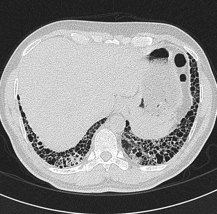
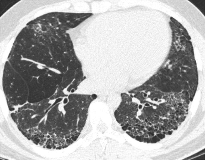
It is well known that high-resolution computed tomography (HRCT) is an essential component of the diagnostic pathway in idiopathic pulmonary fibrosis (IPF). Honeycombing, a common feature of IPF seen on HRCT, is crucial for an accurate diagnosis. Unfortunately, identification of honeycombing is not always straightforward, and there is some disagreement regarding its imaging features. It can be difficult to distinguish honeycombing from traction bronchiectasis and emphysema, although several imaging characteristics can be helpful. Recently, there has been an interest in expanding the use of HRCT beyond diagnosis for disease monitoring and prognostication, and several studies have provided valuable contributions in this regard. Traction bronchiectasis and the extent of fibrosis, for example, have been reported to be powerful prognostic predictors for mortality. Finally, considering the difficulties in diagnosis of "possible usual interstitial pneumonia", clinicians should always be aware that clinical factors must be considered together with HRCT in order to reach an accurate diagnosis and provide appropriate treatment.
©ERS 2014.
Conflict of interest statement
Conflict of interest: Disclosures can be found alongside the online version of this article at
Figures
References
-
- Hunninghake GW, Lynch DA, Galvin JR, et al. . Radiologic findings are strongly associated with a pathologic diagnosis of usual interstitial pneumonia. Chest 2003; 124: 1215–1223. - PubMed
-
- Hansell DM, Bankier AA, MacMahon H, et al. . Fleischner Society: glossary of terms for thoracic imaging. Radiology 2008; 246: 697–722. - PubMed
-
- Johkoh T, Sakai F, Noma S, et al. . Honeycombing on CT: its definitions, pathologic correlation, and future direction of its diagnosis. Eur J Radiol 2014; 83: 27–31. - PubMed
-
- Watadani T, Sakai F, Johkoh T, et al. . Interobserver variability in the CT assessment of honeycombing in the lungs. Radiology 2013; 266: 936–944. - PubMed
Publication types
MeSH terms
LinkOut - more resources
Full Text Sources
Other Literature Sources
Medical