

Case Reports
doi: 10.4103/0301-4738.129786.
A new rosette in retinoblastoma
Affiliations
- PMID: 24881618
- PMCID: PMC4065523
- DOI: 10.4103/0301-4738.129786
Item in Clipboard
Case Reports
A new rosette in retinoblastoma
Indian J Ophthalmol.
2014 May.
Abstract
Retinoblastoma, the most common primary malignant intraocular tumor of childhood is a great success story in pediatric and ocular oncology. Pathology of retinoblastoma is important to guide the treatment modalities. Differentiated retinoblastoma is commonly seen in younger age group. Since a hundred years, we have been observing two typical true rosettes in retinoblastoma in the form of Flexner-Wintersteiner (FW) and Homer Wright (HW) rosettes and in many occasions pseudorosettes have been documented. In the present case report, a third new type of rosette was identified in a differentiated retinoblastoma which had an unusual anterior segment involvement.
Conflict of interest statement
Figures
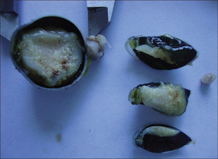
Gross documentation of retinoblastoma specimen. Surface calcification can be noted
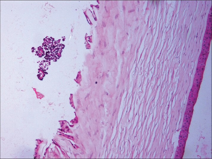
Basophilic tumor cells infiltrating anterior chamber and endothelium of cornea (hematoxylin and esoin (H and E, ×100)
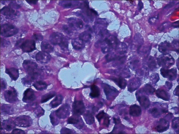
Flexner-Wintersteiner (FW) rosette with clear lumen at the center (H and E, ×400)
Homer Wright (HW) rosette with central tangle of neural filament. There is no clear lumen at the center (H and E, ×400)
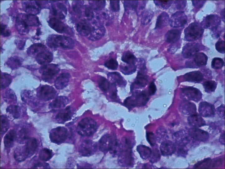
Newly observed rosette with basophilic cuboidal cells occupying the center of lumen. Cytoplasmic extensions of the cells can be noted. (H and E, × 400)
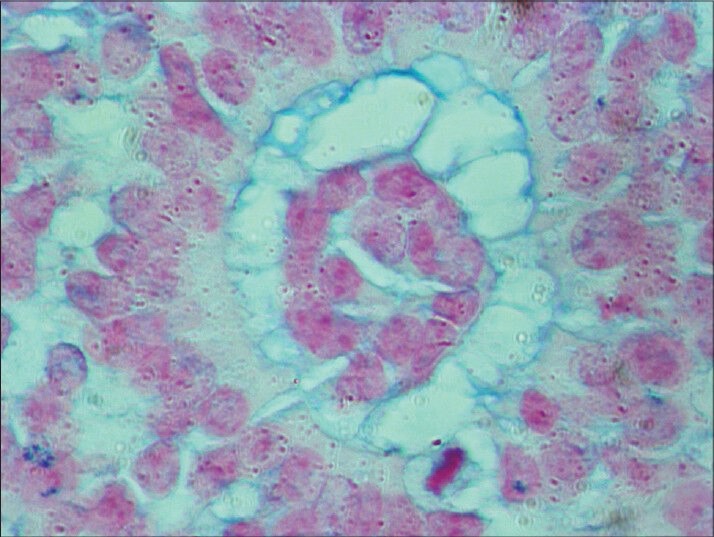
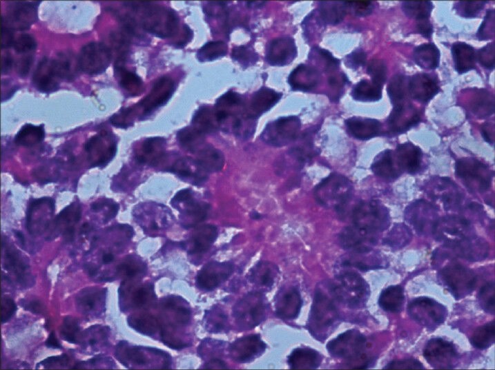
One of the newly observed rosettes stained with alcian blue (HR-AMP). Lumen was stained (×400). HR-AMP = hyaluronidaseresistant acid mucopolysaccharides
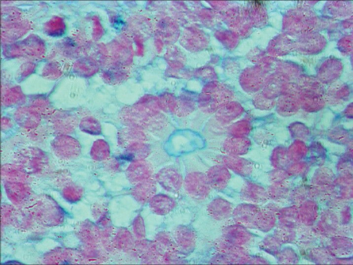
Alcian blue (HR-AMP) stained central lumen of FW rosette in same slide as control (×400)
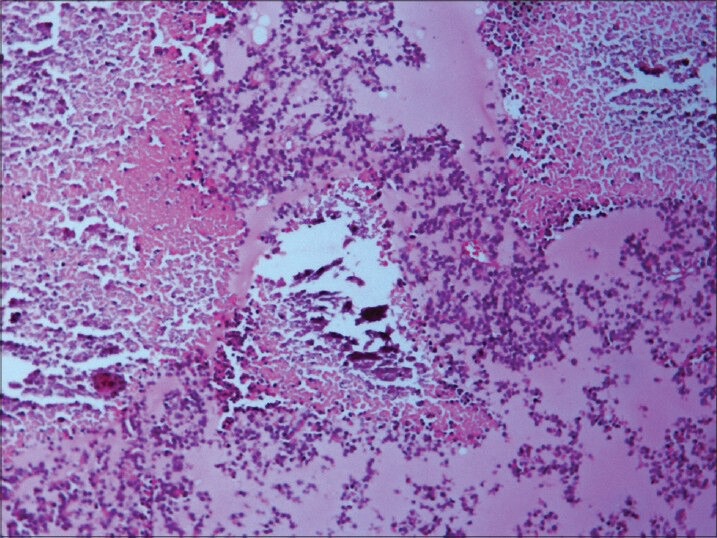
Necrosis and calcification (H and E, ×200)
References
-
- Shields CL, Mashayekhi A, Au AK, Czyc C, Leahey A, Meadow AT, et al. The international classification of retinoblastoma predicts chemoreduction success. Ophthalmology. 2006;113:2276–80. - PubMed
-
- Eagle RC Jr, editor. Eye pathology - An atlas and text. Philadelphia: Lippincott William and Wilkins; 2011.
-
- Tsߣo MO, Zimmerman LE, Fine BS. The nature of retinoblastoma. I. Photoreceptor differentiation: A clinical and histopathological study. Am J Ophthalmol. 1970;69:339–49. - PubMed
-
- Eagle RC., Jr High-risk features and tumour differentiation in retinoblastoma: A retrospective histopathologic study. Arch Pathol Lab Med. 2009;133:1203–9. - PubMed
-
- Madhavan J, Ganesh A, Roy J, Biswas J, Kumaramanickavel G. The relationship between tumour cell differentiation and age at diagnosis in retinoblastoma. J Pediatr Ophthalmol Strabismus. 2008;45:22–5. - PubMed
Publication types
MeSH terms
LinkOut - more resources
Full Text Sources
Other Literature Sources
Medical
