. 2014 Jun 16;24(12):1347-1353.
doi: 10.1016/j.cub.2014.04.044. Epub 2014 May 29.

Role of the primate ventral tegmental area in reinforcement and motivation

Affiliations

Role of the primate ventral tegmental area in reinforcement and motivation

John T Arsenault et al. Curr Biol. .

Abstract

Monkey electrophysiology suggests that the activity of the ventral tegmental area (VTA) helps regulate reinforcement learning and motivated behavior, in part by broadcasting prediction error signals throughout the reward system. However, electrophysiological studies do not allow causal inferences regarding the activity of VTA neurons with respect to these processes because they require artificial manipulation of neuronal firing. Rodent studies fulfilled this requirement by demonstrating that electrical and optogenetic VTA stimulation can induce learning and modulate downstream structures. Still, the primate dopamine system has diverged significantly from that of rodents, exhibiting greatly expanded and uniquely distributed cortical and subcortical innervation patterns. Here, we bridge the gap between rodent perturbation studies and monkey electrophysiology using chronic electrical microstimulation of macaque VTA (VTA-EM). VTA-EM was found to reinforce cue selection in an operant task and to motivate future cue selection using a Pavlovian paradigm. Moreover, by combining VTA-EM with concurrent fMRI, we demonstrated that VTA-EM increased fMRI activity throughout most of the dopaminergic reward system. These results establish a causative role for primate VTA in regulating stimulus-specific reinforcement and motivation as well as in modulating activity throughout the reward system.


Figures

Figure 1
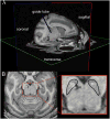
Figure 1. MRI-guided guidetube/electrode implantation
A) Tri-planar cross-section of T1-weighted anatomical image acquired during guide tube insertion. Hypointensity induced by guidetube (see Movie S1) was used to estimate guide tube trajectory/position during surgery (blue cylinder). Estimated VTA target projected from the trajectory of the guidetube (red sphere). B) Post-operative T1-weighted anatomical used to confirm the final electrode position. This transverse slice was the most ventral to exhibit hypointensity from the electrode. The inset displays an expanded view of the midbrain and electrode with the SN outlined. See also Movie S1.
Figure 2
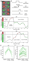
Figure 2. VTA-EM reinforces cue selection (Experiment 1)
A) Four pseudo-randomized, equiprobable trial-types used in the free choice visual cue preference test. New pairs of cues were used in each session. Juice reward probability was equalized across cue position and cue identity. B) Timing schematic of cue presentation, eye movements, juice reward (100 ms, 50 % of trials) and VTA-EM (200 ms, 50% of selections of VTA-associated cue during cue-VTA-EM blocks). Juice and VTA-EM occurred 32–48 ms after cue selection. Cue preference index [(cue B selections − cue A selections)/(cue B selections + cue A selections)] during a single example session of the operant task for subjects M1 (C) and M3 (D). Cue preference index was calculated in bins of 100 and 200 trials for M1 and M3, respectively. Color of data points denote the cue selection followed by VTA-EM on 50% of the trials (gray – no VTA-EM; red – cue B-VTA-EM; green – cue A-VTA-EM). VTA-EM consisted of a 200 ms train of bipolar stimulation pulses (200Hz; 650 μA (M1), 1mA (M3); 2 VTA electrodes stimulated simultaneously). Mean cue preference indices during the 2nd half of each block type for each full session performed by M1 (E) and M3 (F). Green lines denote a session with a consistent trend for increased preference for the cue reinforced with VTA-EM while red lines represent the opposite trend. See also Figure S1 and Table S1.
Figure 3
Figure 3. Pavlovian cue-VTA-EM association motivates future cue selection (Experiment 2)
A) Paradigm consisted of a 20 min. Pavlovian cue-VTA-EM association block surrounded by two cue preference test blocks (no VTA-EM). During the Pavlovian association block, the monkey performed a passive fixation task (0.03 ml juice every 800 – 1200 ms) while only one of the two visual cues (500 ms presentation) shown every 3500 – 6000 ms was temporally associated with VTA-EM (400 ms into cue presentation, bipolar, 200 ms, 200Hz, 1mA, 2 VTA electrodes stimulated simultaneously). The cue preference index from cue preference tests was calculated in bins of 100 trials from single example sessions performed by M2 (B) and M3 (C). Color of data points denotes the preceding Pavlovian association block (gray – no-VTA-EM; red – cue B-VTA-EM; green – cue A-VTA-EM). Mean cue preference index values for each pair of blocks performed by M2 (D) and M3 (E). Green lines denote pairs of cue preference test blocks with a trend for an increased preference of the cue associated with VTA-EM during the intervening Pavlovian association block while red lines represent the opposite trend. See also Figure S2.
Figure 4
Figure 4. fMRI activations induced by VTA-EM (Experiment 3)
Group analysis T-score maps overlaid on coronal slices of the 112 RM-SL T1/T2* anatomical volume (n = 35 runs, M1 = 12 runs, M2 = 5 runs, M3 = 18 runs, fixed effect analysis, VTA-EM – No VTA-EM, FDR corrected, P = 0.001, cluster size 10 voxels). VTA-EM consisted of a 200 ms train of bipolar stimulation pulses (200 Hz, 200 ms, 100 μA – 392 μA, 2 VTA electrodes stimulated simultaneously). Abbreviations: AIP - anterior intraparietal; cnMD - centromedian nucleus; Cd - caudate; DO - dorsal opercular; G - gustatory; GrF - granular frontal; Hc - hippocampus; NA - nucleus accumbens; PAG - periaqueductal gray; Pu - putamen; PrCo - precentral opercular; RN - red nucleus; TPO - temporal parietal occipital; VL - ventral lateral nucleus. See also Figure S3 and Table S2.

References

    1. Bromberg-Martin ES, Matsumoto M, Hikosaka O. Dopamine in motivational control: rewarding, aversive, and alerting. Neuron. 2010;68:815–834. - PMC - PubMed
    1. Schultz W, Dayan P, Montague PR. A neural substrate of prediction and reward. Science. 1997;275:1593–1599. - PubMed
    1. Adamantidis AR, Tsai HC, Boutrel B, Zhang F, Stuber GD, Budygin EA, Tourino C, Bonci A, Deisseroth K, de Lecea L. Optogenetic interrogation of dopaminergic modulation of the multiple phases of reward-seeking behavior. J Neurosci. 2011;31:10829–10835. - PMC - PubMed
    1. Fibiger HC, LePiane FG, Jakubovic A, Phillips AG. The role of dopamine in intracranial self-stimulation of the ventral tegmental area. J Neurosci. 1987;7:3888–3896. - PMC - PubMed
    1. Tsai HC, Zhang F, Adamantidis A, Stuber GD, Bonci A, de Lecea L, Deisseroth K. Phasic firing in dopaminergic neurons is sufficient for behavioral conditioning. Science. 2009;324:1080–1084. - PMC - PubMed

Publication types

LinkOut - more resources