. 2014 Jun 12;7(5):1481-1494.
doi: 10.1016/j.celrep.2014.05.001. Epub 2014 May 29.

Intertissue control of the nucleolus via a myokine-dependent longevity pathway

Affiliations

Intertissue control of the nucleolus via a myokine-dependent longevity pathway

Fabio Demontis et al. Cell Rep. .

Abstract

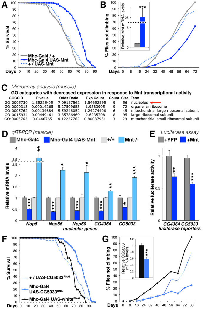
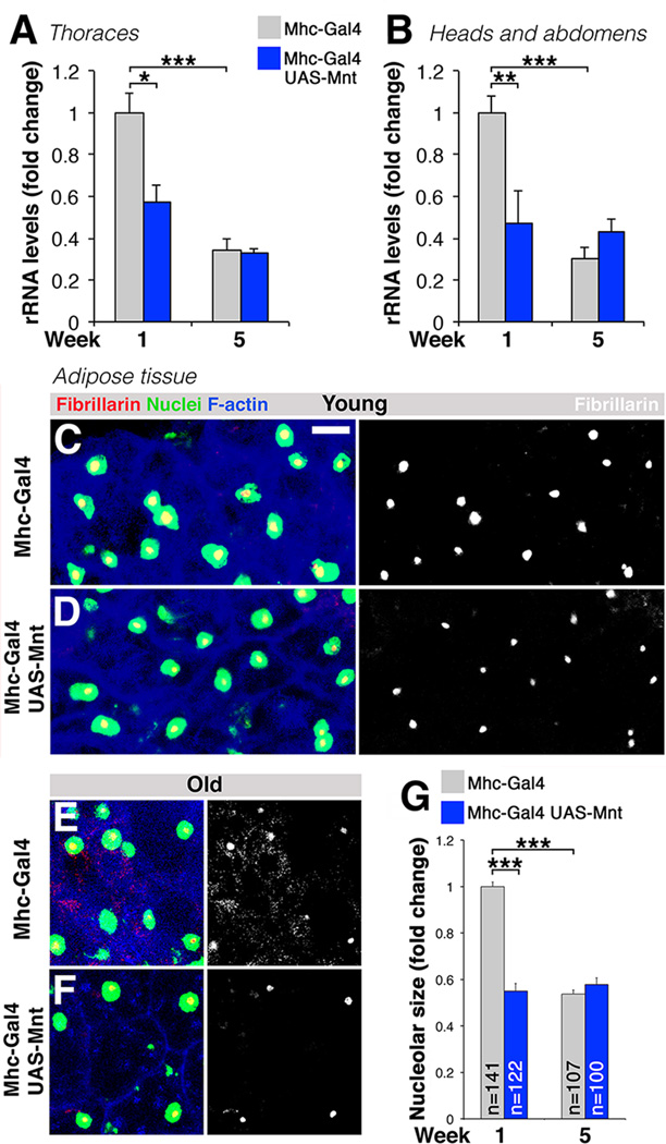
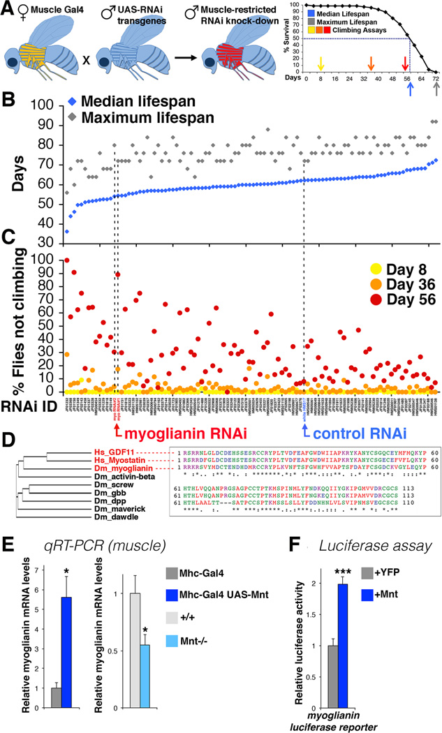
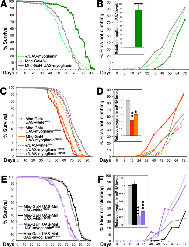
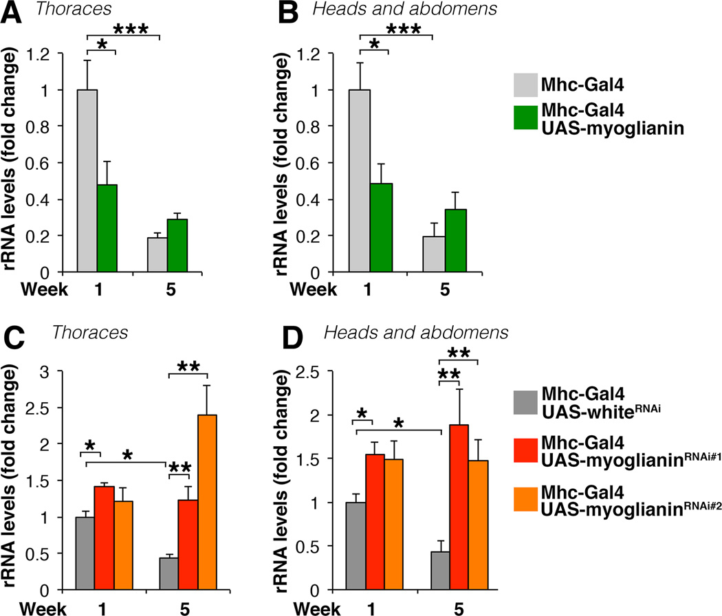
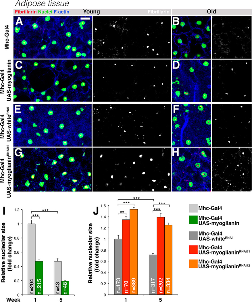
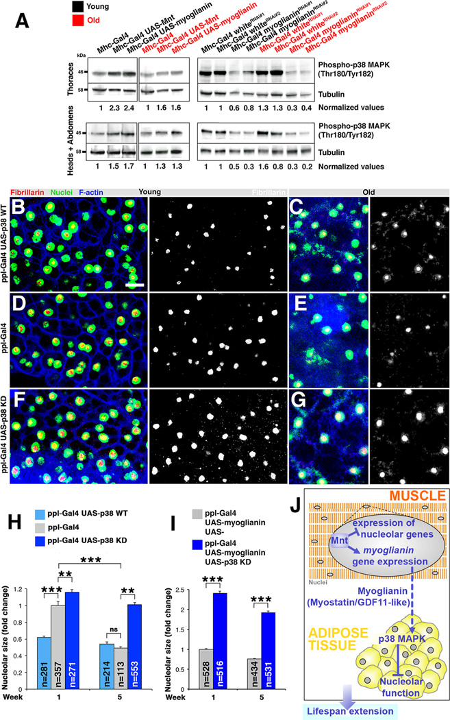
Recent evidence indicates that skeletal muscle influences systemic aging, but little is known about the signaling pathways and muscle-released cytokines (myokines) responsible for this intertissue communication. Here, we show that muscle-specific overexpression of the transcription factor Mnt decreases age-related climbing defects and extends lifespan in Drosophila. Mnt overexpression in muscle autonomously decreases the expression of nucleolar components and systemically decreases rRNA levels and the size of the nucleolus in adipocytes. This nonautonomous control of the nucleolus, a regulator of ribosome biogenesis and lifespan, relies on Myoglianin, a myokine induced by Mnt and orthologous to human GDF11 and Myostatin. Myoglianin overexpression in muscle extends lifespan and decreases nucleolar size in adipocytes by activating p38 mitogen-activated protein kinase (MAPK), whereas Myoglianin RNAi in muscle has converse effects. Altogether, these findings highlight a key role for myokine signaling in the integration of signaling events in muscle and distant tissues during aging.


Figures

Figure 1
Figure 1. Mnt Overexpression in Skeletal Muscle Extends Lifespan and Decreases Age-related Muscle Dysfunction and Nucleolar Protein Gene Expression
(A) Mnt overexpression in skeletal muscle extended the median and maximum lifespan (Mhc-Gal4/UAS-Mnt; blue; n=311; median/maximum lifespan=72.4/90 days; P<0.0001) and reduced the percentage of flies with age-related climbing defects ((B); P<0.001; Log-rank test) as compared with isogenic control siblings (Mhc-Gal/+; grey; n=257; median/maximum lifespan=54.8/78 days) and heterozygous UAS-transgene alone controls (+/UAS-Mnt; blue dotted line; n=236; median/maximum lifespan=55.7/80 days). Relative Mnt mRNA levels are shown in the inset with n=4 and SEM indicated; ***P<0.001; unpaired two-tailed Student’s t-test. (C) Gene expression profiling of skeletal muscle overexpressing Mnt and controls with Affymetrix microarrays. Genes encoding components of the nucleolus and the ribosome are significantly represented among the target genes having decreased expression in response to Mnt transcriptional activity (P<0.05; Exp Count=Expected Count). (D) Relative mRNA levels of nucleolar components from skeletal muscles with increased Mnt levels, from Mnt−/− null flies, and controls. Mnt overexpression (dark blue) decreased the mRNA levels of nucleolar components as compared with controls (dark grey). Conversely, Mnt loss-of-function (blue) led to increased expression. SEM is indicated with n=4; unpaired two-tailed Student’s t-test. (E) Mnt overexpression (blue) decreased the activity of luciferase reporters based on the promoters of the CG4364 and CG5033 genes (which encode nucleolar components) as compared with controls (YFP overexpression; grey). SEM is indicated with n=16. In (D) and (E), *P<0.05; **P<0.01; ***P<0.001; unpaired two-tailed Student’s t-test. (F) CG5033 RNAi in skeletal muscle extended the median and maximum lifespan (Mhc-Gal4/UAS-CG5033RNAi; blue; n=152; median/maximum lifespan=81.9/98; P<0.0001; Log-rank test) and reduced the percentage of flies with age-related climbing defects ((G); P<0.01; Log-rank test) as compared with a mock RNAi treatment against white (Mhc-Gal/UAS-whiteRNAi; black; n=57; median/maximum lifespan=68.6/86) and heterozygous UAS-transgene alone controls (+/UAS-CG5033RNAi; blue dotted line; n=90; median/maximum lifespan=67.7/94). Relative CG5033 mRNA levels are shown in the inset with n=4 and SEM indicated; ***P<0.001; unpaired two-tailed Student’s t-test. See also Figures S1, S2 and S3.
Figure 2
Figure 2. Mnt Overexpression in Muscle Systemically Regulates rRNA Levels and Non-autonomously Reduces Nucleolar Size in Adipocytes
(A–B) qRT-PCR analysis of ribosomal RNA (rRNA) levels in fly thoraces (which consist mostly of skeletal muscle) and heads and abdomens (which consists mostly of non-muscle tissues) from flies overexpressing Mnt specifically in muscle with the Mhc-Gal4 driver. rRNA levels decline during aging in thoraces, heads and abdomens. Flies overexpressing Mnt in skeletal muscle (Mhc-Gal4/UAS-Mnt) display low levels of rRNA already in young age in both thoraces (A) and heads and abdomens (B) in comparison with control flies (Mhc-Gal4/+). SEM and n are indicated with *P<0.05; **P<0.01; ***P<0.001; two-way ANOVA followed by Tukey’s post test. As compared with controls (C, E), overexpression of Mnt in skeletal muscle decreases the nucleolar size of adipocytes already in young age (D), anticipating the decline typically observed in old age (E, F). The nucleolar marker Fibrillarin (red) is shown together with DAPI staining for nuclei (green) and F-actin (phalloidin; blue). In C–F, scale bar is 10 μm. (G) Nucleolar size quantification is shown with SEM and n indicated; *P<0.05; **P<0.01; ***P<0.001; two-way ANOVA followed by Tukey’s post test. See also Figure S4.
Figure 3
Figure 3. Muscle-restricted RNAi Screening Identifies Myoglianin, a Myokine Regulating Lifespan and Induced by Mnt
(A) Scheme of the RNAi screen for myokines regulating lifespan and age-related muscle dysfunction in the fruit fly Drosophila melanogaster. Females carrying the Mhc-Gal4 driver (specific for skeletal muscles) were crossed with males from a collection of Gal4-responsive UAS-RNAi transgenes, generated in a similar genetic background, to knockdown a given cytokine or growth factor specifically in the skeletal muscle of the progeny. Climbing ability (indicative of neuromuscular function) was measured in young (day 8, yellow arrow) and older ages (day 36, orange arrow; and day 56, red arrow), together with the median lifespan (the day at which 50% of flies are alive; blue arrow) and the maximum lifespan (the day at which the last fly in a cohort is alive; grey arrow). (B) RNAi screen results ordered by ascending median lifespan. Each blue dot represents the median lifespan (indicated in days on the y axis) of a cohort of typically 60 flies with a muscle-specific RNAi intervention (TRiP RNAi ID is shown on the x axis of the graph in (C), see also Figure S5). Corresponding grey dots represent the maximum lifespan of the same cohort. Two different RNAi treatments in skeletal muscles against the TGF-β ligand myoglianin (arrow and name in red) shortened the median lifespan as compared with a control RNAi against white (arrow and name in blue). (C) The percentage of flies with climbing defects at day 8 (yellow), 36 (orange), and 56 (red) for the cohorts of flies with lifespan reported in (B). Myoglianin RNAi increased the percentage of flies with climbing defects in old age as compared with a control RNAi against white. Note that none of the RNAi treatments resulted in climbing defects in young age (day 8), indicating that the differences observed in older age are not due to developmental defects. See Extended Experimental Procedures and Figure S5 for statistical analysis of the screen. (D) Phylogenetic analysis of Drosophila BMP and TGF-β ligands shows that Myoglianin is the sole Drosophila homolog of the highly related human TGF-β ligands Myostatin (GDF8) and GDF11. Alignment of the mature Drosophila Myoglianin and human GDF11 and Myostatin peptides shows extensive sequence identities (47%; *) and similarities (69%; :). (E) Mnt overexpression in skeletal muscles significantly increased myoglianin expression, whereas muscles from Mnt−/− null flies had lower myoglianin mRNA levels. SEM is indicated with n=4; *P<0.05; unpaired two-tailed Student’s t-test (F) Overexpression of Mnt (blue) in Drosophila S2R+ cells resulted in higher myoglianin-luciferase reporter activity than controls (YFP overexpression; grey). SEM is indicated with n=16; ***P<0.001; unpaired two-tailed Student’s t-test.
Figure 4
Figure 4. Mnt/Myoglianin Signaling Regulates Lifespan and Muscle Function during Aging
(A) Myoglianin overexpression (Mhc-Gal4/UAS-myoglianin; green; n=458; median/maximum lifespan=75.5/94 days) in skeletal muscles extended the median and maximum lifespan (P<0.0001; Log-rank test) and reduced the percentage of flies with age-related climbing defects ((B); P<0.01; Log-rank test) as compared with isogenic control siblings (MhcGal4/+; grey; n=152; median/maximum lifespan=59.3/78 days) and heterozygous UAS-transgene alone controls (UAS-myoglianin/+; green dotted line; n=370; median/maximum lifespan=53/86 days). (C) Muscle-specific knockdown of myoglianin (RNAi#1: red; n=275; median/maximum lifespan=55.9/88 days; RNAi#2: orange; n=731; median/maximum lifespan=59.1/86 days) shortened the median lifespan (P<0.0001; Log-rank test), as compared with white knockdown (grey, control; n=290; median/maximum lifespan=69.5/96 days) and heterozygous flies carrying UAS-RNAi transgenes alone (UAS-myoglianinRNA#1/+: red dotted line, n=367; median/maximum lifespan=67.2/92 days; UAS-myoglianinRNAi#2/+: orange dotted line, n=323; median/maximum lifespan=67.1/92; and UAS-whiteRNAi/+: grey dotted line, n=307; median/maximum lifespan=69.6/88). (D) The percentage of flies with climbing defects progressively increased during aging in controls but to a greater extent in flies with myoglianin knockdown in muscles (P<0.01; Log-rank test). (E) Myoglianin knockdown (RNAi#1: pink, n=142; median/maximum lifespan=66.5/88 days; and RNAi#2: violet, n=192; median/maximum lifespan=66.6/88 days) reduced the lifespan extension resulting from Mnt overexpression in muscles (P<0.001; Log-rank test) as compared with control white knockdowns (RNAi#1: grey, n=140; median/maximum lifespan= 78.2/100 days; and RNAi#2, black, n=179; median/maximum lifespan=82.4/104). (F) A higher percentage of flies with climbing defects was observed during aging when myoglianin levels were reduced in skeletal muscles overexpressing Mnt (P<0.01; Log-rank test). In A–F, relative myoglianin mRNA levels are shown in insets with n=4 and SEM indicated; *P<0.05, **P<0.01, and ***P<0.001; unpaired two-tailed Student’s t-test. See also Figures S3 and S6.
Figure 5
Figure 5. Muscle-derived Myoglianin Regulates Nucleolar Function during Aging
(A–D) qRT-PCR analysis of rRNA levels in fly thoraces (which consist mostly of skeletal muscle) and heads and abdomens (which consists mostly of non-muscle tissues) from flies expressing different levels of myoglianin in skeletal muscle. (A–B) rRNA levels decline during aging in thoraces, heads and abdomens. Flies overexpressing myoglianin in skeletal muscle (Mhc-Gal4/UAS-myoglianin) display low levels of rRNA already in young age in both thoraces and heads and abdomens in comparison with control flies (Mhc-Gal4/+). (C–D) rRNA levels do not decline during aging in thoraces, heads and abdomens from flies with myoglianinRNAi in skeletal muscle (Mhc-Gal4/UAS-myoglianinRNAi) in comparison with control flies (Mhc-Gal4/UAS-whiteRNAi). In (A–D), n=4 and SEM is indicated; *P<0.05, **P<0.01, and ***P<0.001; two-way ANOVA followed by Tukey’s post test.
Figure 6
Figure 6. Muscle-derived Myoglianin Regulates Nucleolar Size in Adipocytes
(A–H) Immunostaining of adipose tissue for the nucleolus (Fibrillarin; red), F-actin (phalloidin; blue), and nuclei (DAPI; green). (C, D) Overexpression of myoglianin in skeletal muscles decreased the nucleolar size of adipocytes already in young age as compared with controls (A), anticipating the decline in nucleolar size typically observed in old age (B). (G, H) Muscle-restricted myoglianin RNAi increased nucleolar size in adipocytes in young and old age, in comparison with a control RNAi against white (E, F). In A–H, scale bar is 10 μm. Nucleolar size quantification is shown in (I, J) with SEM and n indicated; **P<0.01; ***P<0.001; two-way ANOVA followed by Tukey’s post test.
Figure 7
Figure 7. Mnt Activity in Muscle Remotely Activates p38 MAPK via Myoglianin to Regulate Nucleolar Size in Adipocytes
(A) Western blot analysis of fractions from either thoraces (consisting mainly of muscles; upper panels) or heads and abdomens (consisting mainly of non-muscle tissues; lower panels) from controls and flies overexpressing Mnt and myoglianin (left panels) or with RNAi-mediated knockdown of myoglianin or white (right panels) in skeletal muscles from young (1 week) and old (8 weeks) flies. Mnt and Myoglianin increase the levels of active phospho-p38 MAPK in both muscle and non-muscle tissues as compared with controls (left panels) whereas myoglianin knockdown had converse effects (right panels). Relative quantification of phospho-p38 MAPK levels normalized to α-Tubulin is indicated. See also Figure S7. (B–H) Adipocytes labeled with the nucleolar marker Fibrillarin (red), phalloidin (F-actin; blue), and DAPI (nuclei; green). (B) In young age (1 week), wild-type (WT) p38 MAPK overexpression in adipocytes with the ppl-Gal4 driver led to significantly smaller nucleoli than in controls (D, H), leading to a premature decline in nucleolar size which is otherwise normally observed in old age (C, E, H). Conversely, bigger nucleoli were observed in young (F) and old age (G, H) after overexpression of dominant negative, kinase-dead (KD) p38 MAPK. (I) Similar results were obtained also when myoglianin was concomitantly overexpressed with p38 KD (ppl-Gal4/UAS-p38KD;UAS-myoglianin/+;), in comparison with overexpression of a UAS-myoglianin transgene together with an empty UAS- that drives no transgene expression and which is used to control for Gal4 titration effects in the different genotypes analyzed (ppl-Gal4/UAS-; UAS-myoglianin/+;). Scale bar is 10 μm. Statistical analysis was done with two-way ANOVA followed by Tukey’s post test in (H–I). In (H–I), n is indicated with SEM. **P<0.01; ***P<0.001; ns=not significant. (J) Summary scheme. Muscle-specific overexpression of Mnt extends lifespan and prevents age-related muscle functional decay. In the nuclei (grey) of myofibers (orange), the transcription factor Mnt reduces rRNA levels and the expression of components of the nucleolus, a regulator of aging. In addition, Mnt increases the expression of the TGF-beta ligand myoglianin, the Drosophila ortholog of human Myostatin and GDF11. In turn, muscle-specific overexpression of myoglianin extends lifespan and reduces rRNA levels in the whole organism and nucleolar size in adipocytes (yellow) by activating p38 MAPK, a known transducer of TGF-beta signaling, while myoglianin RNAi in muscle has converse effects. In addition to inducing systemic effects, Myoglianin acts in an autocrine manner to reduce rRNA levels in muscle and preserves muscle function during aging. These findings highlight that signaling events in skeletal muscle can influence systemic aging and lifespan via muscle-derived secreted proteins, i.e. myokines. Myokine-based coordination of nucleolar function across aging tissues may represent an assurance mechanism for robust lifespan determination in aging organisms.

Comment in

  • GDF11/myostatin and aging.
    Patel VK, Demontis F. Patel VK, et al. Aging (Albany NY). 2014 May;6(5):351-2. doi: 10.18632/aging.100666. Aging (Albany NY). 2014. PMID: 24902527 Free PMC article. No abstract available.

References

    1. Amthor H, Macharia R, Navarrete R, Schuelke M, Brown SC, Otto A, Voit T, Muntoni F, Vrbova G, Partridge T, et al. Lack of myostatin results in excessive muscle growth but impaired force generation. Proc Natl Acad Sci U S A. 2007;104:1835–1840. - PMC - PubMed
    1. Awasaki T, Huang Y, O'Connor MB, Lee T. Glia instruct developmental neuronal remodeling through TGF-beta signaling. Nat Neurosci. 2011;14:821–823. - PMC - PubMed
    1. Bai H, Kang P, Hernandez AM, Tatar M. Activin signaling targeted by insulin/dFOXO regulates aging and muscle proteostasis in Drosophila. PLoS Genet. 2013;9:e1003941. - PMC - PubMed
    1. Bostrom P, Wu J, Jedrychowski MP, Korde A, Ye L, Lo JC, Rasbach KA, Bostrom EA, Choi JH, Long JZ, et al. A PGC1-alpha-dependent myokine that drives brown-fat-like development of white fat and thermogenesis. Nature. 2012;481:463–468. - PMC - PubMed
    1. Boulon S, Westman BJ, Hutten S, Boisvert FM, Lamond AI. The nucleolus under stress. Mol Cell. 2010;40:216–227. - PMC - PubMed

Publication types

MeSH terms

Substances

Associated data